
Figures & data
Figure 1. Follow-up.
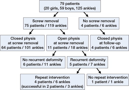
Table 1. Group assignment
Figure 2. Lateral distal tibial angle (LDTA). This is determined by measuring the angle created by the intersection of the central axis of the tibia and a second line drawn across the epiphyseal surface of the distal tibia.
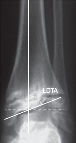
Table 2. Results of radiographic analyses
Figure 3. Case series. Full correction within 26 months in a boy with a meningomyelocele. The deformity was balanced at the time of physeal closure
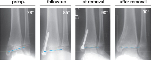
Table 3. Literature review