Figures & data
Figure 1. Flow chart of patient selection.
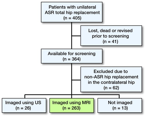
Figure 3. Box plot showing median WB Cr levels, interquartile range (boxes), and 95% CIs (whiskers) with outliers (circles), presented according to grade of g. medius atrophy.
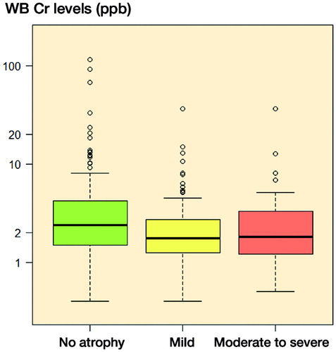
Table 1. Clinical and radiological details of the patients
Table 2. Prevalence (%) of fatty atrophy in each of the gluteal muscles
Figure 4. Distribution of grade of atrophy according to different pseudotumor (PT) findings.
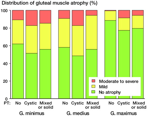
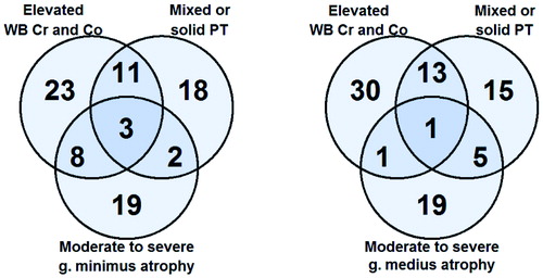
Figure 5. Venn diagrams showing the overlapping of findings of interest in patients with moderate-to-severe g. minimus atrophy (left panel) and with moderate-to-severe g. medius atrophy (right panel). Patients with moderate-to-severe g. maximus atrophy are not shown because of the very low prevalence.