
Figures & data
Figure 1. Measurement of the maximum diameter of the ossific nuclei.
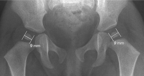
Figure 2. A. A girl with fragmentation of the left ossific nucleus (grade-1 AVN) at 12-month follow-up. Treatment had been initiated at 2 days of age for left-sided instability. B. Normal appearance of the femoral head at 3 years. C. Normal hip development at final check-up. The observation time was 5 years 7 months.
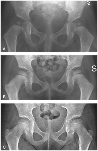
Figure 3. A. A boy with mottled appearance of the left ossific nucleus (grade-1 AVN) at 12-month follow-up. Treatment had been initiated at 2 days of age for left-sided dislocation (Barlow-positive). B. Normal appearance of the femoral head at 8 years.
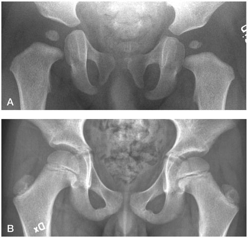
Figure 4. Flow chart detailing inclusion of patients in the study. Assessment of AVN was performed by review of 1-year and subsequent radiographs (Kalamchi-MacEwen grading), hospital files, and a diagnosis registry to a mean observation time of 6.5 years.
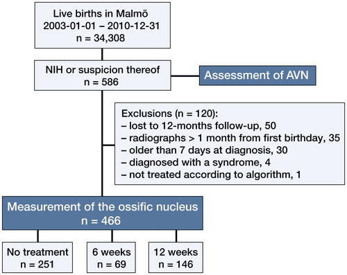
Figure 5. A higher degree of neonatal instability was associated with a smaller ossific nucleus at 12 months. Treatment differed between these groups (red dots).
Hips with a positive Ortolani test (i.e. dislocated in resting position) had smaller ossific nuclei than hips with a positive Barlow test (i.e. resting in the acetabulum but dislocatable) at 12 months. All these hips had 12 weeks of treatment (blue dots, subgroup analysis of dislocated hips). (n denotes the number of hips in each group. Error bars represent the 95% CI of the mean).
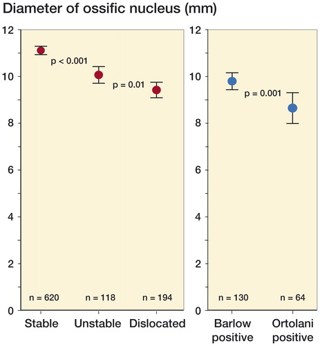
Table. 1 Comparison of the mean size of the ossific nucleus between study groups