Figures & data
Figure 1. (A) Coronary angiography showing diffuse atherosclerosis in RCA with site of plaque rupture denoted by small arrowheads (>). (B) Grey-scale IVUS showing plaque rupture at proximal PDA (inset). (C) VH assessment of mid-distal RCA showing large amount of necrotic core (inset). (D) Coronary angiography of RCA after POBA of PDA.
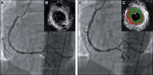
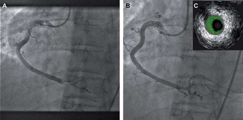
Figure 2. (A) Coronary angiography showing thrombotic occlusion in distal RCA, (B) Coronary angiography of RCA after aspiration thrombectomy. (C) VH assessment of distal RCA showing presence of thrombus with minimal necrotic core (inset).