Figures & data
Figure 1. EKG on admission showing electrocardiographic signs of right-ventricular overload, i.e. vertical-to-right axis, right bundle branch block.
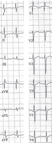
Figure 2. Transthoracic echocardiographic 4-chamber view showing an enlarged right ventricle (RV), right atrium (RA) and a thrombus (arrow) crossing the interatrial septum.
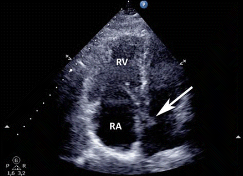
Figure 3. Transesophageal echocardiographic short-axis view showing the right atrium (RA), left atrium (LA), aorta (AO) and thrombus through the foramen ovale (arrow).
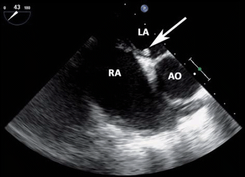
Figure 4. Transesophageal 3-dimensional echocardiographic view through the roof of the left-(LA) and right atrium (RA) looking down on the mitral valve (MV) and the thrombus (arrow).
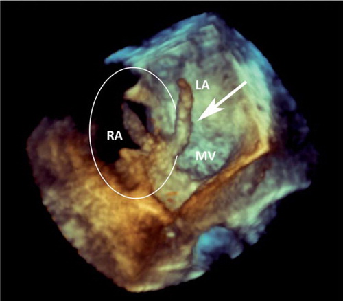
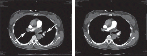
Figure 5. Contrast-enhanced CT scans of the chest showing adjacent slices with bilateral thrombi (arrows).