
Figures & data
Figure 1. Diagram of the ‘stent jail’ technique: A. Proximal stenting using the jailed wire. B. Proximal stenting using the non-jailed wire.
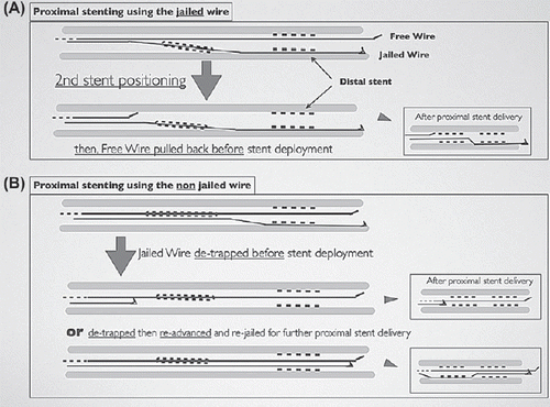
Table I. Clinical and procedural characteristics.
Figure 2. A. LAO view: severe multifocal right coronary artery disease, the artery being very calcified and tortuous. The distal lesion was the most severe. B. Jailing the buddy wire during distal stenting. Note the actual shape of the 5F guide catheter in a ‘power position’. C. More proximal stent positioning over the jailed wire (JW), the free wire has been pulled back (FW). D. Final result.
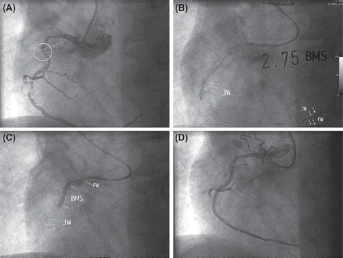
Table II. Solutions for difficult PCI.
Table III. Proximal versus distal buddy-in-jail techniques.