Figures & data
Figure 1. Two-dimensional transthoracic echocardiography image. Four-chamber apical view indicative of mass in the left atrium (white arrow).
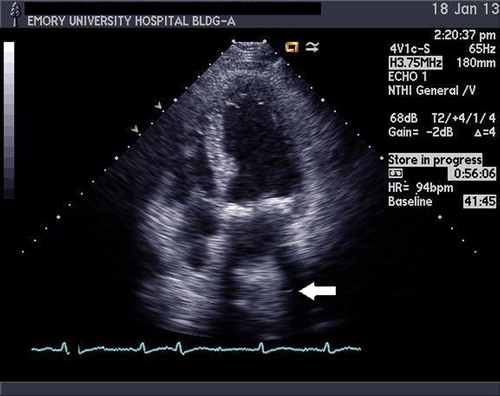
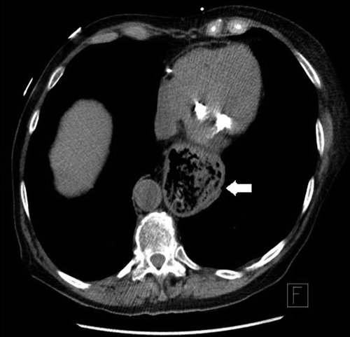
Figure 2. Chest computed tomography axial plane image showing a large hiatal hernia compressing the heart, especially on the left atrium (white arrow).