
Figures & data
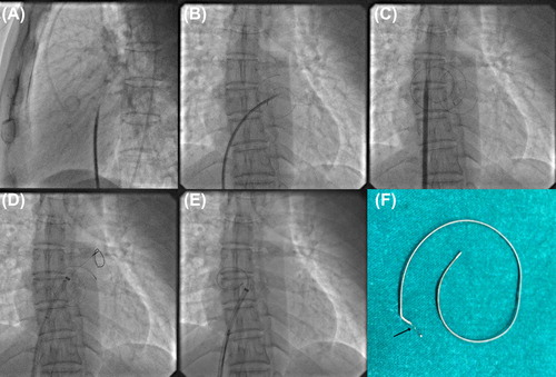
Figure 1. Transseptal puncture using Brokenbrough needle and Mullin's dilator (A). Broken 0.025 inch coiled tip guidewire in left atrium (B). Septal dilation with 14F septal dilator after introduction of another 0.025 inch coiled tip guidewire through 8F Mullin's dilator (C). Introduction of gooseneck snare in left atrium through 10F Amplatzer long delivery sheath to capture broken guidewire (D) and pulling it into sheath (E). Severed proximal end of broken guidewire after successful retrieval (arrow, F).
Supplemental material