Figures & data
Table 1. Clinical details of the patients and newborns with preeclampsia, HELLP syndrome, IUGR, and the normal control group of well characterized group of patients [Mylonas et al. 2006a; 2006b; Schiessl et al. 2005; 2006]. Data represent mean ± standard deviation.
Figure 1. Immunohistochemical staining reaction of inhibin-βC in placental syncytiotrophoblast cells. Syncytiotrophoblast cells demonstrated a positive cytoplasmatic staining intensity for inhibin-βC antibody in normal ( A; lens 20, top left panel), preeclamptic ( B; lens 40, top right panel), HELLP (C; lens 40, lower left panel), and IUGR (D; lens 40, lower right panel) placental tissue.
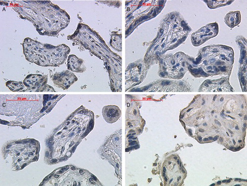
Figure 2. Immunohistochemical staining reaction of inhibin-βC in placental extravillous trophoblast cells. Extravillous trophoblast cells demonstrated a positive cytoplasmatic staining intensity for inhibin-βC antibody in normal (A; lens 20, upper left panel), preeclamptic (B; lens 20, upper right panel), HELLP (C; lens 20, lower left panel), and IUGR (D; lens 40, lower right panel) placental tissue.

Figure 3. Immunohistochemical evaluation of the inhibin-βC subunits in normal, preeclamptic, and HELLP placenta tissue. The immunoreactive score for inhibin-βC did not show any significant differences between normal, preeclamptic, HELLP, or IUGR placental tissue in syncytiotrophoblast cells and extravillous trophoblast. Data represent mean ± SEM. Significance was assumed at p < 0.05.