Figures & data
Figure 1. IL-6 expression in normal peritoneal and adhesion fibroblasts. Adhesion fibroblasts had increased IL-6 expression as compared to normal peritoneal fibroblasts. Exposure to hypoxia increased IL-6 expression in both normal and adhesion fibroblasts. (*, ** p < 0.05 as compared to normal peritoneal fibroblasts, *** p < 0.05 as compared to untreated adhesion fibroblasts and compared to hypoxia treated normal peritoneal fibroblasts).
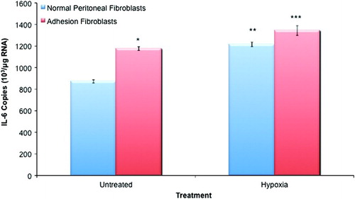
Figure 2. TNF-α expression in normal peritoneal and adhesion fibroblasts. Adhesion fibroblasts had increased TNF-α expression as compared to normal peritoneal fibroblasts. Exposure to hypoxia increased TNF-α expression in both normal and adhesion fibroblasts. (*, ** p < 0.05 as compared to normal peritoneal fibroblasts, *** p < 0.05 as compared to untreated adhesion fibroblasts and compared to hypoxia treated normal peritoneal fibroblasts).
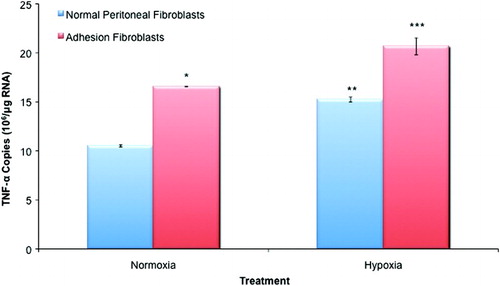
Figure 3. The adhesion phenotype. The adhesion phenotype can be induced during in vitro culture of human peritoneal fibroblasts by exposure to hypoxia. When compared to normal peritoneal fibroblasts, these changes include increases in interferon (IFN)-γ, TGF-β1, ECM markers including type I and III collagens as well as fibronectin, COX2, VEGF, iNOS, and decreases in TGF-β3, MMPs, the ratio of tPA to PAI-1, apoptosis, and NO.
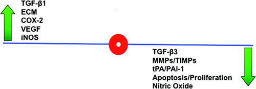
Table 1. Human oligonucleotide primers for β-actin, IL-6, and TNF-α.