Figures & data
Figure 1. High magnification by an inverted microscope equipped with Nomarski differential interference contrast optics and video system. Arrows indicate nuclear vacuoles observed through a 100x (1.40 numerical aperture) objective lens. Dotted lines indicate the width of the sperm head (3.3 µM) and the maximum diameter of a nuclear vacuole (2.5 µM).
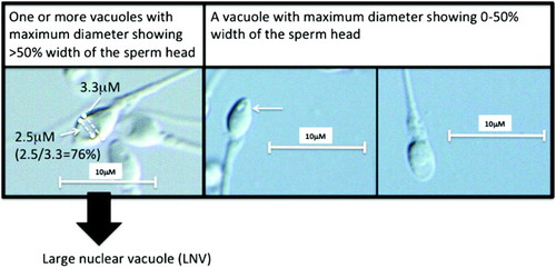
Table 1. Background of the patients.
Table 2. Comparison of the % LNV between patients with idiopathic male infertility and varicocele.
Table 3. Correlation between the % LNV and the parameters of the conventional semen analysis and SMAS.