Figures & data
Figure 1. Three-dimensional (3D) imaging of spermatozoon.Four different angles of a representative spermatozoon nucleus. For each cell, the following was measured: 1) the volume of the nucleus, 2) the volume of pan-centromeric signals, 3) the volume of the chromosome 17 centromere specific signal, 4) the number of observed chromocenter(s) (6 chromocenters for the depicted cell), 5) the distance from the tail attachment (reference point A) to the centromere signal (4.1 µm for the depicted cell), and 6) the long axis (8.4µm), (distance point from A to point B). Fluorescence in situ hybridization (FISH) probes: human chromosome pan-centromeric – green; α-satellite centromere specific chromosome 17 - red. Measurements from the tail attachment point to the centromere of chromosome 17.
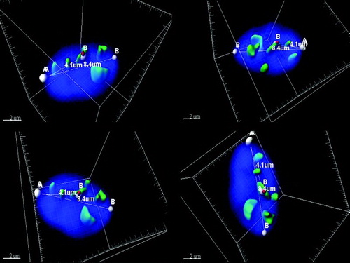
Figure 2. The schematic representation of the sperm nucleus. A) Radial cell division: P - peripheral, C – central (equally divided). B) Longitudinal cell division: A - acrosomal region, M - medial region, T - tail region (equally divided).
Figure 3. Descriptive evaluation of three variables in the motile and immotile spermatozoa. Three variables were taken into account (the number of chromocenters, the radial as well as the longitudinal positions of the centromere of chromosome 17 within the nucleus) to assess the variations of unique prototypes of spermatozoa. Localization of the centromere of chromosome 17: the radial position described as periphery (P) or central (C); the longitudinal position classified as acrosomal (A), medial (M), or tail region (T) and the numeral number represents the quantity of chromocenters in the sperm nucleus. Example: PA1: centromere of chromosome 17 located in the periphery (outer) acrosomal part of the nuclei with 1 observed chromocenter.
Table 1. Compression between motile and immotile spermatozoa.