Figures & data
Figure 1. Postnatal body weight development of male offspring exposed to fluoxetine (5 mg/kg, n = 10; 10 mg/kg, n = 8; 20 mg/kg, n = 10; and control, n = 10) via placenta and lactation.
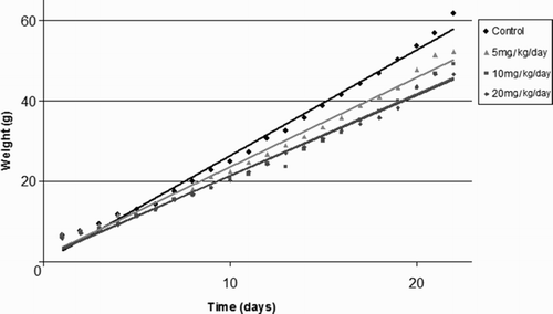
Table 1. Body, testis, and epididymis weight of 22-day old male offspring exposed to fluoxetine (5 mg/kg, n = 10; 10 mg/kg, n = 8; 20 mg/kg, n = 10; and control, n = 10) via placenta and lactation.
Table 2. Volume of different testicular components of 22-day old male offspring exposed to fluoxetine (5 mg/kg, n = 10; 10 mg/kg, n = 8; 20 mg/kg, n = 10; and control, n = 10) via placenta and lactation.
Figure 2. Photomicrographs of testicular sections of 22 day old offspring exposed to fluoxetine via placenta and lactation. A) Testicular section of control animal, luminated seminiferous tubules (arrow). B) Testicular section of control rat, luminated seminiferous tubules (ST) and Sertoli cell (arrow). C) Testicular section from fluoxetine 5 mg/kg group, luminated seminiferous tubules (arrow) and intertubular compartment (star). D) Testicular section from fluoxetine 10 mg/kg group, note the germ cells showing morphological characteristics of apoptosis (black arrows) in a seminiferous tubule (ST), Leydig cells (white arrows), and lymphatic space (star). E) Testicular section from fluoxetine 20 mg/kg group, note the presence of luminated seminiferous tubules (white arrows) and tubules undergoing lumen formation (black arrows). F) Testicular section from fluoxetine 20 mg/kg group, note the presence of seminiferous tubules undergoing lumen formation (black arrow) and a luminated seminiferous tubules (white arrows) in greater magnification.
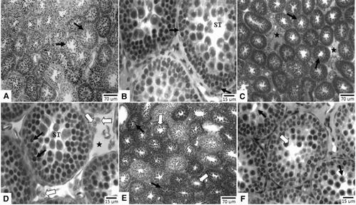
Table 3. Morphometric data and Sertoli cell number of 22-day old male offspring exposed to fluoxetine (5 mg/kg, n = 10; 10 mg/kg, n = 8; 20 mg/kg, n = 10; and control, n = 10) via placenta and lactation.
Figure 3. Plasma testosterone levels (ng/mL) of 22 day old offspring exposed to fluoxetine (Flu; 5 mg/kg, n = 10; 10 mg/kg, n = 8; 20 mg/kg, n = 10; and control, n = 10) via placenta and lactation. Results were expressed as mean and (±) standard deviation.
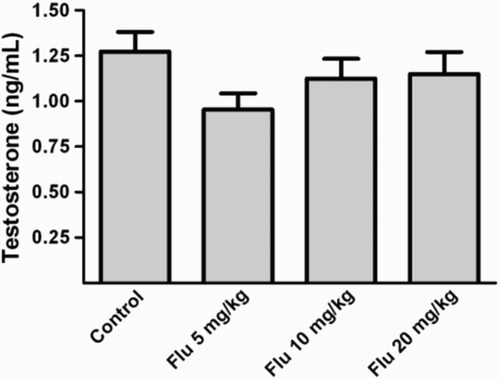
Table 4. Leydig cell cellular, nuclear, and cytoplasmatic area of 22-day old male offspring exposed to fluoxetine (5 mg/kg, n = 10; 10 mg/kg, n = 8; 20 mg/kg, n = 10; and control, n = 10) via placenta and lactation.