Figures & data
Figure 1. Example of live cells images taken in an inverted microscope after individual cell isolation by micromanipulation. SC: Sertoli cells; SGA: spermatogonia: type-1 spermatogonia elongated and fusiform (arrows) and type-2 spermatogonia elongated (white arrowhead) and round (black arrowhead); ST1: primary spermatocytes; CD34+: umbilical cord blood CD34+ cells. Bars: = 20 μm (SC, SGA, ST1) and = 10 µm (CD34+)
Figure 2. Reverse transcriptase-polymerase chain reaction characterization of OCT4, KIT, ITGA6, ITGB1, and CD3D in human testicular and hematopoietic cells. The subsequent code was employed to assign testicular and hematopoietic cells: SG: spermatogonia; ST1: primary spermatocytes; SC: Sertoli cells; DGC: diploid germ cell suspensions; W: whole-testicular cell suspensions; CD34+: umbilical cord blood CD34+ cells; Ly: lymphocytes. MW indicates the molecular weight marker of 100bp (Invitrogen). No bands were detected in the negative control (N). Human beta actin (ACTB) was used as house-keeping gene. OCT4: octamer-binding transcription factor 4; KIT: v-Kit Hardy-Zuckerman 4 Feline Sarcoma Viral Oncogene Homolog; ITGA6: integrin alpha 6; ITGB1: integrin beta 1; CD3D: CD3 delta
Figure 3. Immunocytochemical localization of OCT4, KIT, ITGA6, and ITGB1 in adult human testicular cells and hematopoietic cells. The subsequent code was employed to assign testicular and hematopoietic cells: SG: spermatogonia; ST1: primary spermatocytes; SC: Sertoli cells; CD34+: umbilical cord blood CD34+ cells; Ly: lymphocytes. Replacement of primary antibody with phosphate buffered saline (PBS) in Ly provided the negative controls. OCT4: octamer-binding transcription factor 4; KIT: v-Kit Hardy-Zuckerman 4 Feline Sarcoma Viral Oncogene Homolog; ITGA6: integrin alpha 6; ITGB1: integrin beta 1
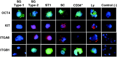
Figure 4. Immunohistochemical localization of cell markers in paraffin sections of adult human testis. A, B inset-section without hematoxylin counterstaining) OCT4; C, inset) KIT; D) ITGA6; E, inset) ITGB1; and F) example of a negative control with primary antibody omission. BL: basal lamina; PTC: peritubular cells; SC: Sertoli cells; SG: spermatogonia; Ad: spermatogonia A-dark; Ap: spermatogonia A-pale; ST1: primary spermatocytes; Sa: round spermatids; Sd: elongated spermatids; Sz: spermatozoa; De: scaled cells in the lumen of the seminiferous tubule; Lu: lumen of the seminiferous tubule. Scale bars = 5 µm (B, C, E), 10 µm (D, F), and 20 µm (A).
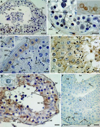
Table 1. Marker positivity (+) in germ cells and Sertoli cells.
Table 2. Forward and reverse primers used for RT-PCR.