Figures & data
Figure 1. Comparison of serum anti-sperm antibody levels in control, sham-control, TESA, and MESA groups. Each bar indicates mean absorbance on ELISA (optical density at 414 nm) and the error bars represent the mean ± SEM of six animals. Significant at ***(P < 0.001). Data indicates no significant difference in the serum antisperm antibodies levels between the control and sham-control groups. The titer in TESA group was significantly elevated in comparison with MESA or control animals (P < 0.001). However, in MESA group titer was higher than control and sham-control animals; but the differences were not statistically significant. TESA: testicular sperm aspiration; MESA: microsurgical epididymal sperm aspiration
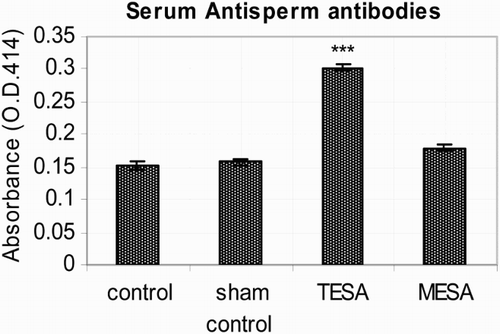
Table 1. Tabulated values of weight and volume of the testis taken from control and other experimental groups.
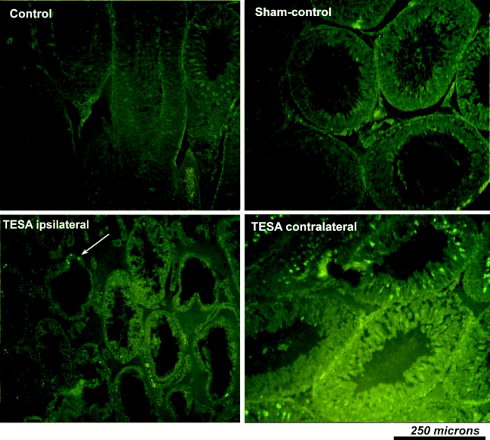
Figure 2. Photomicrograph of testis stained with hematoxylin and eosin taken from various experimental groups. Photomicrograph from control and sham-control rat testes demonstrating normal pattern of spermatogenesis. Histological picture from both ipsi and contralateral testes indicates alteration in TESA and MESA group. However, TESA group testis showing more damage especially ipsilateral testis was severely damaged and devoid of sperm cells in the majority of the tubules after TESA procedure. TESA: testicular sperm aspiration; MESA: microsurgical epididymal sperm aspiration
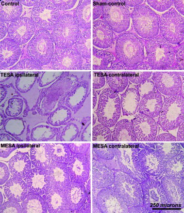
Figure 3. Histomorphometric analysis (relative values) of testicular components in control and experimental groups (A, B, C, D). Data showed significant increase in the number of tubular cross sections (numerical density) per unit area in the TESA testes (A) this could be due to the reduction in seminiferous tubular diameter as indicated by data shown in B, along with loss of epithelial height (C). D) indicates the reduction in the volume of various testicular components (tubules, epithelium, and connective tissues), these findings correspond to the significant decrease in testicular weight and volume of the gross measurements in corresponding groups. These data indicate a significant loss of germinal epithelium in both TESA and MESA with a significant damage in TESA ispsilateral testis. Each bar represents mean ± SEM of six animals. Significant at **(P < 0.01) and ***(P < 0.001). TESA: testicular sperm aspiration; MESA: microsurgical epididymal sperm aspiration
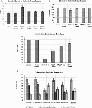
Figure 4. Histomorphometric values (relative values) of epididymis in control and experimental groups are shown in A, B, C, D, E, F (height of the epithelium, diameter of tubules, number of tubules, and volume of various epididymal components in caput, corpus, and cauda region, respectively). Histometric data indicates that apart from the change in epithelium (E) and connective tissue (F) in MESA-ipsilateral epididymis, there was no obvious change in the rest of the groups. Each bar represents mean ± SEM of six animals. Significant at *** (P < 0.001); TESA: testicular sperm aspiration; MESA: microsurgical epididymal sperm aspiration
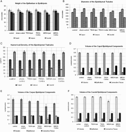
Figure 5. Photomicrograph of testis taken from various experimental groups after staining with TUNEL technique to demonstrate apoptotic positive cells. No obvious apoptotic cells seen in control and sham-control. However, increased apoptosis of germ cells was seen in TESA testis (predominantly in the regions of germ/sperm cells). Though it appears that the number of apoptotic positive cells were relatively reduced in TESA ipsilateral testis when compared to other groups and contralateral side, actually it was because of the loss of seminiferous epithelia and sperm cells in majority of the tubules after TESA. TESA: testicular sperm aspiration; MESA: microsurgical epididymal sperm aspiration