
Figures & data
Figure 1. High-magnification on an inverted microscope equipped with Nomarski differential interference contrast optics and a video system. The arrows indicate nuclear vacuoles observed through a 60× (1.42 numerical aperture) objective lens. (A) Spermatozoa with a nuclear vacuole exhibiting a >50% transverse diameter of the sperm nuclear area. The dotted lines indicate the width of the sperm head (3.1 μm) and the maximum diameter of a nuclear vacuole (1.8 μm). (B) Spermatozoa with a nuclear vacuole exhibiting a ∼50% transverse diameter of the sperm nuclear vacuole. (C) Spermatozoa without any nuclear vacuoles in the sperm nuclear area.
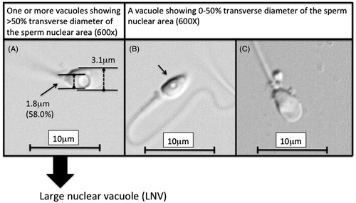
Table 1. Patient characteristics.
Table 2. Comparison of the 1st and 2nd ejaculates at evaluation for male infertility.
Table 3. Changes in the sperm nuclear vacuoles among the patients who underwent varicocele repair.
Table 4. The sperm nuclear vacuoles among the patients who underwent medicinal therapy.