Figures & data
Figure 1. Example for a probeset used for fluorescence in situ hybridization (FISH) in the present study on a normal diploid female metaphase (A) and in a human sperm with a small supernumerary marker chromosome (sSMC) 15. (A) All probe sets used were designed as shown exemplarily for combination of chromosome-6- and -15-specific probeset. Here this probeset was hybridized on a normal diploid female, peripheral blood derived metaphase spread: partial chromosome paints (pcp) for short (p) and long (q) arm identify chromosome (= #) 6; a combination of an alphoid centromeric probe for 15p11.1 to 15q11.1 and a satellite III probe for 15p11.2 was applied to stain chromosome 15. (B) An example of how 3D-FISH analyses looked like using Cell-P-program (Olympus) is shown. The same sperm is shown twice. In the upper lane only chromosome 15 and sSMC(15) are depicted; as only probes covering overall 15p11.2 to 15q11.1 were applied the normal chromosome 15 shows a smaller staining than the sSMC(15) and both can be distinguished. In the lower lane all color channels used are depicted, i.e., the sSMC(15) and chromosomes 15 as well as 6 can be identified. Chromosomes 6, 15, and sSMC(15) were positioned in periphery. Chromosome 15 is located towards the sperm tail, chromosome 6 towards the head. Chromosome 6 and sSMC(15) are localized in close proximity, but also chromosome 15 and sSMC(15) are located together and not on opposite sides of the nucleus.
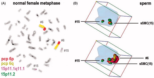
Figure 2. Positioning of the studied chromosomes, including small supernumerary marker chromosome (sSMC) 15, with respect to localization towards sperm center or periphery in both studied brothers and normal person. Positioning of chromosomes 6, 15, 18, 19, 21, X and Y, and the sSMC(15) towards sperm center and periphery in fertile brother (F) and infertile brother (I) studied, here compared to normal situation (N) according to [Manvelyan et al. Citation2008b]. Experiments were done using 3D-FISH (fluorescence in situ hybridization) as depicted in .
![Figure 2. Positioning of the studied chromosomes, including small supernumerary marker chromosome (sSMC) 15, with respect to localization towards sperm center or periphery in both studied brothers and normal person. Positioning of chromosomes 6, 15, 18, 19, 21, X and Y, and the sSMC(15) towards sperm center and periphery in fertile brother (F) and infertile brother (I) studied, here compared to normal situation (N) according to [Manvelyan et al. Citation2008b]. Experiments were done using 3D-FISH (fluorescence in situ hybridization) as depicted in Figure 1.](/cms/asset/299f2311-e053-4343-af17-d3df2866c3c0/iaan_a_979956_f0002_c.jpg)
Figure 3. Positioning of the studied chromosomes, including small supernumerary marker chromosome (sSMC) 15, with respect to localization towards sperm head, middle, or tail in both studied brothers and normal person. Depicted is the positioning of chromosomes as in towards sperm head, middle part, and tail in fertile brother (F) and infertile brother (I) studied here and compared to normal situation (N) according to [Manvelyan et al. Citation2008b]. Experiments were done using 3D-FISH (fluorescence in situ hybridization) as depicted in .
![Figure 3. Positioning of the studied chromosomes, including small supernumerary marker chromosome (sSMC) 15, with respect to localization towards sperm head, middle, or tail in both studied brothers and normal person. Depicted is the positioning of chromosomes as in Figure 2 towards sperm head, middle part, and tail in fertile brother (F) and infertile brother (I) studied here and compared to normal situation (N) according to [Manvelyan et al. Citation2008b]. Experiments were done using 3D-FISH (fluorescence in situ hybridization) as depicted in Figure 1.](/cms/asset/50069a5e-1bbe-466e-a43d-e2eb7da7f67b/iaan_a_979956_f0003_c.jpg)
Figure 4. Colocalization of the small supernumerary marker chromosome (sSMC) 15 and the corresponding tested chromosomes in both brothers. Percentage of sperm cells with colocalization of the sSMC and the corresponding tested chromosomes as in in fertile brother (F) and infertile brother (I). Experiments were done using 3D-FISH (fluorescence in situ hybridization) as depicted in .
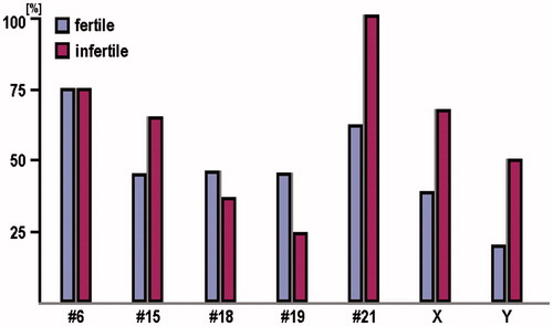
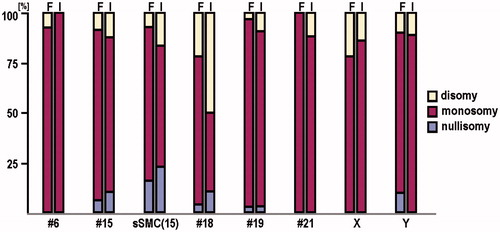
Figure 5. Monosomy and disomy for the studied chromosomes and the small supernumerary marker chromosome (sSMC) 15 in both brothers. Percentage of sperm with monosomy and disomy for the studied chromosomes and the sSMC(15) in fertile brother (F) and infertile brother (I). Experiments were done using 3D-FISH (fluorescence in situ hybridization) as depicted in .