Figures & data
Table 1. Comparison of semen parameters and male age in fertile individuals and infertile men with varicocele.
Figure 1. Comparison of the mean percentages of 2′, 7′-dichlorodihydrofluorescein (DCFH+), TUNEL+, chromomycin A3 (), and intensity of DCFH (I-DCFH) between individuals with varicocele and fertile individuals. *p < 0.05; **p < 0.01.
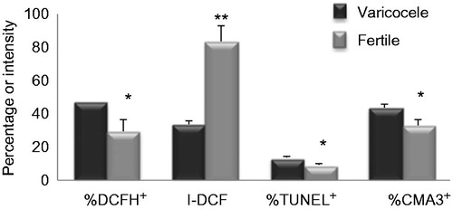
Figure 2. Comparison of mean percentage of DNA methylation and intensity of DNA methylation between individuals with varicocele and fertile individuals. % GDM: % global DNA methylation; Int-GDM: intensity of DNA methylation; FCM: flow cytometry analyses. *p < 0.05.
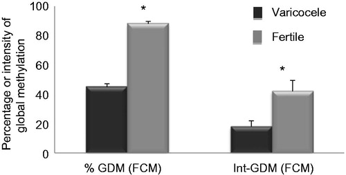
Figure 3. Fluorescence images of sperm stained for DNA methylation (A), DNA fragmentation (B), and protamine deficiency (C). DNA methylation was assessed by antibody against 5-methylcytosine. DNA fragmentation was assessed by TUNEL staining (spermatozoa with fragmented DNA stained green (TUNEL positive, shown with arrow), whereas TUNEL negative sperm appear as red due to propidium iodide staining of nuclei). Protamine deficiency was assessed by chromomycin A3 (CMA3) staining, spermatozoa with bright yellow staining were considered as protamine deficient or CMA3 positive (arrow head), while spermatozoa with dull yellow staining were considered as having a normal amount of protamine or CMA3-negative.
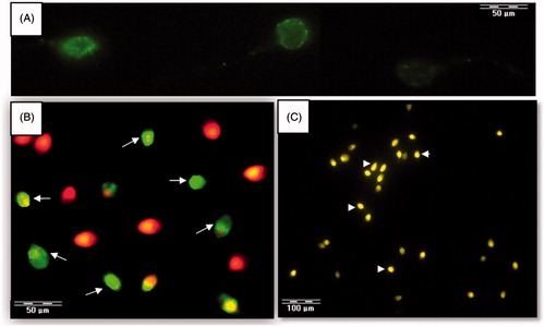
Table 2. Correlation between percentages of sperm positive for TUNEL, chromomycin A3 (CMA3), 2′, 7′-dichlorodihydrofluorescein (DCFH), and intensity of DCFH (I-DCFH) with sperm parameters in individuals with grades II and III varicocele and fertile individuals.
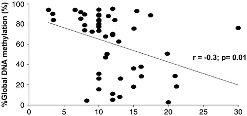
Figure 4. Relationship between percentages of sperm global DNA methylation (GDM) with sperm DNA fragmentation in individuals with grades II and III varicocele and fertile men. N = 59.