Figures & data
Figure 1. MRI brain scan from the patient with concurrent Fabry’s disease and amyotrophic lateral sclerosis. (A and B) T2 weighted transverse MRI scan showing symmetrical white matter change; (C) Sagittal FLAIR MRI scan showing white matter changes in the cerebral hemispheres; and (D) T2 weighted transverse MRI showing white matter changes in the brainstem.
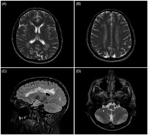
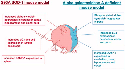
Figure 2. Autophagy is dysregulated in mouse models of both Fabry’s disease and SOD1-G93A amyotrophic lateral sclerosis.