Figures & data
Figure 1. a) Size analysis, b) SEM and c) TEM images of AO-loaded nanoparticles.
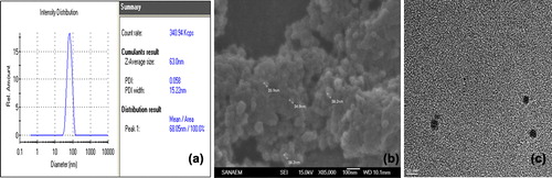
Figure 2. AFM image of AO-loaded PHEMA nanoparticles.
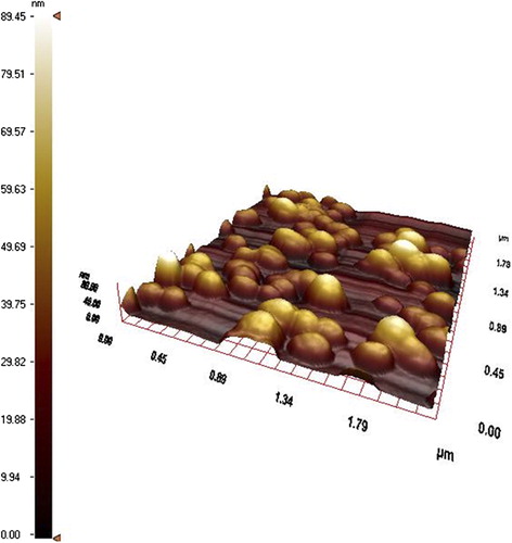
Figure 3. FTIR spectra of a) AO, b) AO-loaded PHEMA nanoparticles c) PHEMA nanoparticles.
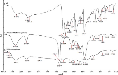
Figure 4. Fluoresence microscopy images of L929 cells. a) Transmission and b) fluoresence after 24 h incubation at 37°C with AO-loaded PHEMA nanoparticles. Different labeling of cell membranes or compartments can be noticed indicating the release of the AO has occured from polymeric nanoparticles.
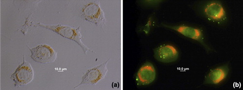
Figure 5. The effect of AO-loaded PHEMA nanoparticle concentration on the cell viability of L929 mouse fibroblast cells.
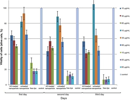
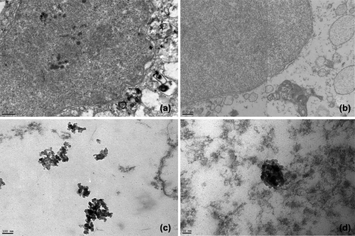
Figure 6. TEM images of L929 cells incubated for 24 h with AO-loaded PHEMA nanoparticles showing coagulation of nanoparticles in cell compartments at an accelerating voltage of 120 kV (a, c, d). L929 cells just as control is seen in b).