Figures & data
Figure 1. Visual observation of B. licheniformis cell lysate supernatant after 24 h of incubation in test tubes. (a) Control sample having only bacterial cell lysate supernatant; (b) Cell lysate supernatant with aqueous solution of HAuCl4 showing gold precipitation (color changed to pink).
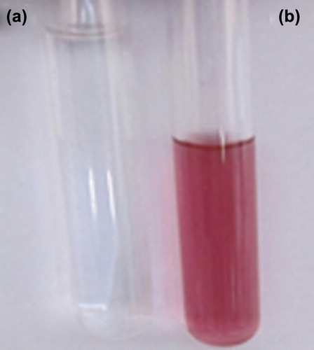
Figure 2. The optical absorption spectra of gold nanoparticle at different time intervals (a) Absorbance of gold nanoparticles against wavelength (control, 12h, 24h, and 48h); (b) The graph shows relationship between optical absorbance of gold nanoparticles with incubation time; (c) Stability of gold nanoparticles at room temperature (1 month and 2 month).
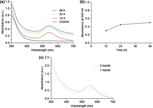
Table I. Optical properties, size, and zeta potential of synthesized gold nanoparticles from cell lysate supernatant of B. licheniformis at different time interval.
Figure 3. Histogram showing particle size distribution of gold nanoparticles.
Figure 4. SEM and EDX analysis (a) representative SEM micrograph of gold nanoparticles synthesized by B. licheniformis cell lysate supernatant; (b) Spot EDX profile confirming the presence of gold in sample.
Figure 5. Antimicrobial activity of AuNPs against (a) E. coli, (b) P. aeruginosa, (c) B. Subtilis. Cup containing (I) sterile deionized water as control and (II) gold nanoparticles; concentration of AuNPs in each plate was 25 μg/mL.
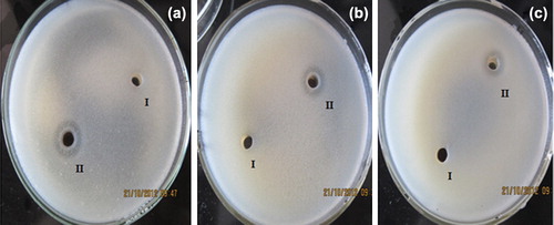
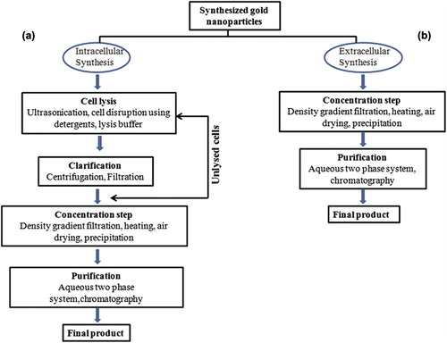
Figure 6. A comparative flow chart of downstream processing steps to get purified gold nanoparticles from (a) Intracellular synthesis, (b) Extracellular synthesis (CLS mediated).