Figures & data
Figure 1. Operating microscope images (A, C, E, 5.5×) displaying the gross anatomy of the surgical procedures and the corresponding schematic drawings (B, D, F). A and B demonstrate the surgical procedures of repairing the femoral nerve by epineurium neurorrhaphy. C and D demonstrate the surgical procedures for repairing the femoral nerve by small gap sleeve bridging with a biodegradable chitin conduit. The 6 mm biodegradable chitin conduits were placed at the repair site and a 2 mm gap was maintained between the proximal and distal nerve segments. E and F demonstrate the removal of the sensory axons by ablating the right L2–4 DRGs and the arrows in photo E indicate the right L2–4 DRGs.
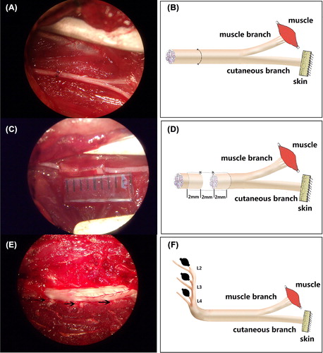
Figure 2. Light microscopy images (10 × 100) of the parent motor axons in the proximal stumps (A: Group I; B: Group II), the regenerating axons in the distal stumps (C: Group I; D: Group II), and the misrouted motor axons in the cutaneous stumps (E: Group I; F: Group II). The scale bar is 20 μm.
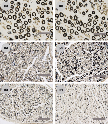
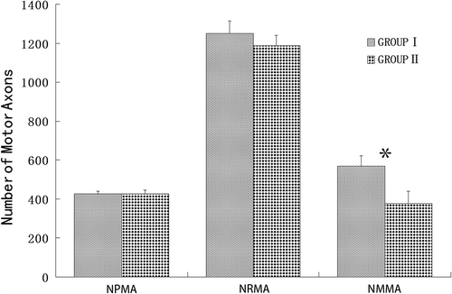
Figure 3. A comparison of the different numbers of parent motor axons in the proximal stump and regenerating motor axons in the distal stump and saphenous nerve stump for Group I and Group II. NPMA: the number of parent motor axons; NRMA: the number of regenerating motor axons; NMMA: the number of misrouting motor axons. Neither the number of parent motor axons nor the regenerating motor axons exhibited a significant difference between Group I and Group II. The number of misrouting motor axons in the saphenous nerve for Group I is significantly more than Group II.