Figures & data
Figure 1. Representative 785-nm spectra after baseline correction and smoothing for low, medium, and high methemoglobin concentrations extracted from the oxidation experiment with different volumes of 10% potassium ferricyanide (power at the sample, 10 mW).
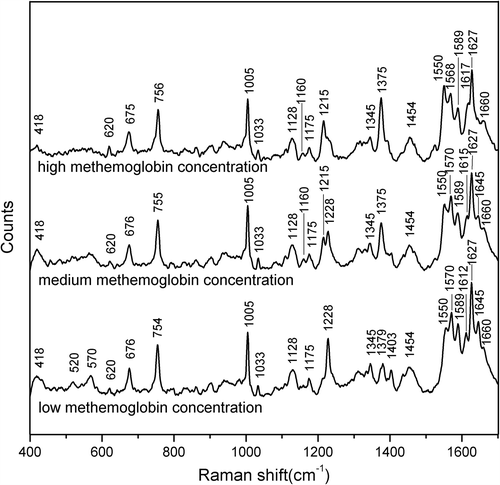
Figure 2. Comparison of the spectra recorded for the Hb solutions with (a) low methemoglobin concentration and (b) high methemoglobin concentration using 514.5-nm and 785-nm laser excitation, showing the major band assignments in the range of 500–2000 cm− 1.
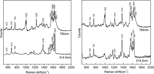
Figure 3. Two-dimensional Raman correlation synchronous spectra in the range of 1200–1700 cm− 1 with the perturbation of oxidation by potassium ferricyanide.
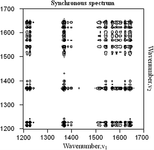
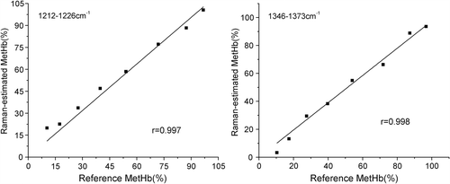
Figure 4. Methemoglobin concentration values estimated from Raman spectroscopy and measured using spectrophotometry. Each point represents one measurement at a given methemoglobin concentration (MetHb(%)). Each panel presents the 8 measurements used to calculate each least squares regression line. The correlation coefficients (r) were all statistically significant. Raman-estimated methemoglobin concentration was calculated using the regression coefficients obtained from the formula (a) in the ranges of 1210–1230 cm− 1 and the 1340–1380 cm− 1. The spectrophotometrically measured methemoglobin concentration is shown on the horizontal axis.