Figures & data
Figure 1. Nanofibrous scaffold designed by electrospinning method. A) Electrospinning set, B) Nanofibrous film and C) Collagen-coated nanofibrous film.
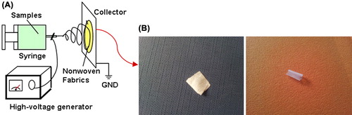
Figure 2. The grafting process of scaffolds loaded with stem cells on damaged skin.
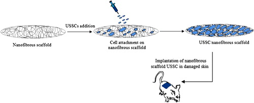
Figure 3. SEM images of the un-cross linked nanofibrous PHBV mat (A: 5000× – B: 10000×) and the collagen-coated nanofibrous PHBV mat (C: 5000× – D: 10000×).
Table I. The mechanical and physical properties of electrospun nanofibrous PHBV mats.
Figure 4. Percentage of original wound area after 21 days for the control, the un-modified nanofibrous scaffold without USSCs, the un-modified nanofibrous scaffold with USSCs, the collagen-coated nanofibrous scaffold without USSCs, and the collagen-coated nanofibrous scaffold with USSCs.
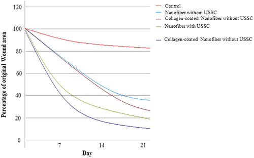
Figure 5. Histology of wounds by H&E staining in the different groups on post-operative day 21. A) The nanofibrous PHBV scaffold without USSCs. B) The nanofibrous PHBV scaffold with USSCs. C) The collagen-coated nanofibrous PHBV scaffold without USSCs. D) The collagen-coated nanofibrous PHBV scaffold with USSCs. E) The normal skin. F) control. (Abbreviations: FD fibrinous debris; EP – epithelialization; HF – hair follicle; HB – hair bulb; SB – sebaceous gland; VS – vascularized section). Scale bars: 200 m.
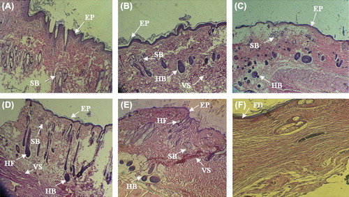
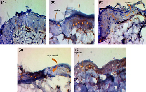
Figure 6. Immunostaining or epidermal markers expression (cytokeratin-10) on day 21. A) The un-modified nanofibrous scaffold without USSCs. B) The un-modified nanofibrous scaffold with USSCs. C) The collagen-coated nanofibrous scaffold without USSCs. D) The collagen-coated nanofibrous scaffold with USSCs. E) The normal skin. Scale bars: 50 μm.