Figures & data
Figure 1. Illustration of surgical procedures. 1/2 Proximal tibial nerve segment was served as father nerve to repair the distal nerve stump using biodegradable chitin conduits with a gap of 1 mm.
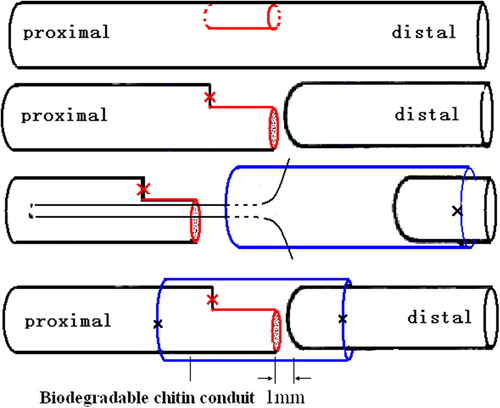
Table I. Comparison of myelinated axon numbers for all groups.
Figure 2. Electrophysiological recordings of the compound muscle action potential of gastrocnemius muscles 4 months after operation. The motor NCV of the tibial nerve in the sham control group was 49.5 ± 4.9 m/s (A) while the value of the distal tibial nerve segment in the experimental group was 27.8 ± 5.3 m/s (B).
Figure 3. Histological sections through the reconstructed nerves 4 months after surgery. A: The proximal part of the tibial nerve (normal tibial nerve). B: The distal part of the tibial nerve was repaired with 1/2 number of proximal donor axons (regenerated tibial nerve).
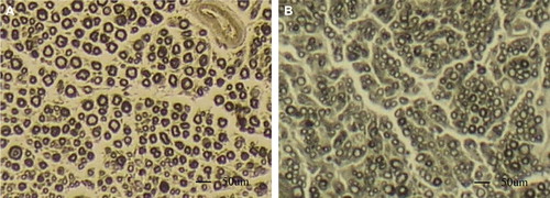
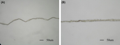
Figure 4. Myelinated nerve fiber isolated from the tibial nerve. A: single fiber isolated from the tibial nerve in sham control group; B: Regenerated fiber at the site of suture with biodegradable chitin conduits. A myelinated axon sprouts collateral branches at the node of Ranvier. (Observed with light microscopy working at 100× magnification).