Figures & data
Figure 1. The upper row – a photograph of P3HB filling material (I), porous 3D implants: life-size (II), and × 10 magnified (III); the lower row – SEM images of P3HB porous implant.
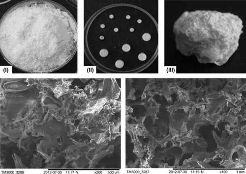
Table I. Characterization of P3HB-based 3D implants.
Figure 2. Cross section samples of the bones at the defect site; implants: I – P3HB, II – P3HB/HA composite (HA 20 wt%), III – Bio-Oss® (arrows).
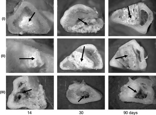
Figure 3. Radiological data on the regeneration dynamics of the model defects of bone tissue in experiments with different implants.
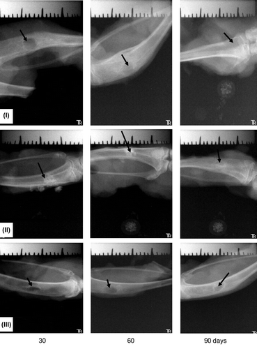
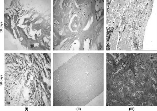
Figure 4. New tissue at the implantation site: I – P3HB, II – P3HB/HA, III – Bio-Oss – at different time points after surgery. Magnification: × 100 hematoxylin–eosin.
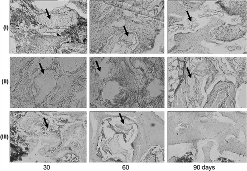
Figure 5. Radiological data on the regeneration dynamics of the defects of bone tissue infected with Staphylococcus aureus in experiments with different implants: I – bone allograft; II – powdered P3HB; III – powdered P3HB/tienam.
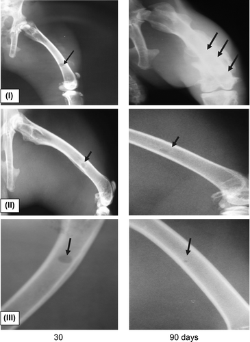
Table II. Results of microbiological investigation of the samples.
Figure 6. New tissue at the site of implantation of I – powdered bone allograft, II – P3HB, and III – P3HB/tienam at different time points after surgery. Magnification: × 100 hematoxylin–eosin.