
Figures & data
Figure 1. The nanofibrous mat designed by electrospinning method. (A) The electrospinning set, (B) The nanofibrous PHBV mat, (C) The chitosan-crosslinked nanofibrous PHBV scaffold, and (D) The rolling of nanofibrous mat using a thermal agent.
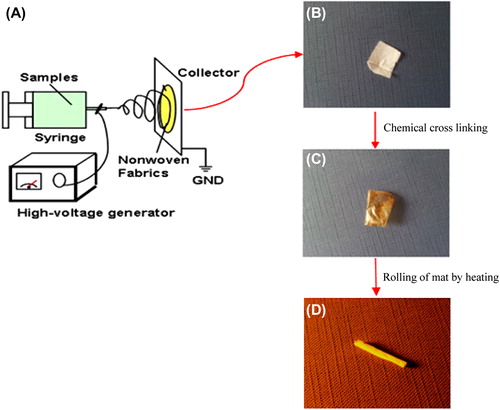
Figure 2. SEM images of the un-crosslinked nanofibrous PHBV mat (A: 5000×, B: 20000×), and the chitosan-crosslinked nanofibrous PHBV mat (C: 5000×, D: 20000×).
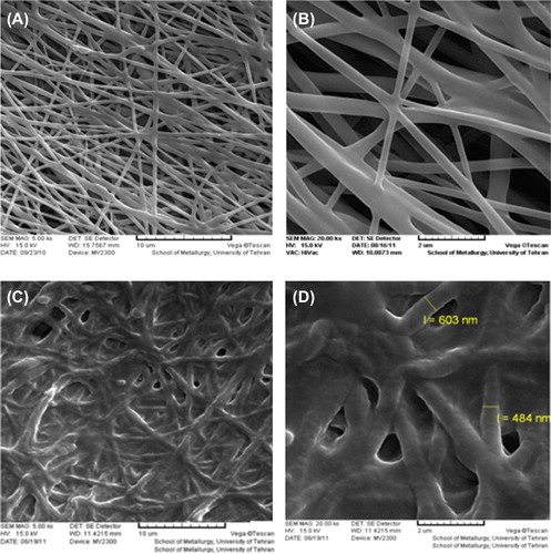
Figure 3. SEM images of the chitosan-crosslinked nanofibrous conduit. (A) The tubular conduit (45×), (B) The nanofibrous structure of designed conduit (20000×), (C) The diameter of tube wall (1000×).
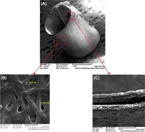
Table I. The mechanical and physical properties of elecrospun PHBV mats.
Figure 4. Cell culture on the nanofibrous mats, and the control. (A) The control (TCPS), (B) The un-modified nanofibrous PHBV mat, (C) The chitosan-crosslinked nanofibrous mat.
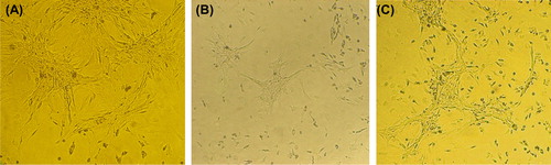
Figure 5. SEM images of cultured Schwann cells on the nanofibrous mats. (A) The un-modified nanofibrous PHBV mat, (B) The chitosan-crosslinked nanofibrous mat (Mag: 2000×).
Table II. Results of MTT assay for the un-crosslinked, and the chitosan-crosslinked nanofibrous PHBV mats.