Figures & data
Table I. Primer sequences used in real-time qPCR.
Figure 1. Morphology of rat cardiac stem cell and vascular endothelial cells. (A) Rat primary c-kit+ cardiac stem cells after purification through FCM. (B) Vascular endothelial differentiation of rat c-kit+ cardiac stem cells, cells converted from fusiform and triangular to roundness in shapes after day 6.
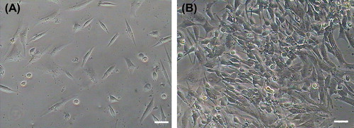
Figure 2. Detection of rat c-kit+ cardiac stem cells markers by immunofluorescence staining. The results show that rat c-kit+ cardiac stem cells at various passages were positive for the c-kit, GATA 4, Nkx2.5 and Flk 1 cell surface markers but cTnT and CD31 were weakly expressed.
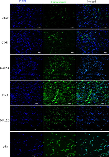
Figure 3. Identification surface markers of vascular endothelial cells, the expression of CD31 (FITC, green) and CD144 (Cy5, red) were detected using immunofluorescence.
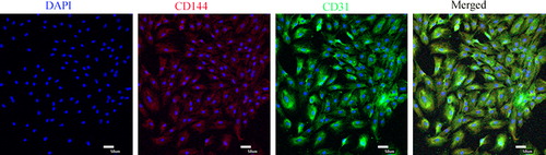
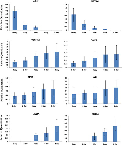
Figure 4. Real time qPCR analyses of PI3K/Akt cell signaling pathway members and vascular endothelial cell markers in endothelial cell differentiation. The result indicated that after endothelial cell induction the specific genes, including PI3K/Akt cell signaling pathway members (PI3K and Akt), and endothelial cell markers (CD31, VEGFR-2 and CD144) were detected, and gene expression level showed a time-lapse increase, cardiac stem cells markers (c-kit and GATA 4) showed a time-lapse gradually decrease.