Figures & data
Figure 1. P. deceptionensis DC5 on TSA medium (a), P. deceptionensis DC5 on TSA with 1 mM AgNO3 (b), control flask (c) after incubation period, culture supernatant of P. deceptionensis DC5 containing nanoparticles (d), respectively.
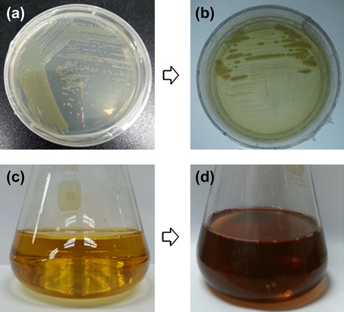
Figure 2. Time dependent UV-Vis spectra of reaction mixture, which shows the synthesis of silver nanoparticles (a), XRD pattern of silver nanoparticles (b).
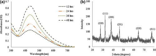
Figure 3. FE-TEM image of silver nanoparticles (a–d), EDX spectra of silver nanoparticles (e), elemental mapping of silver nanoparticles; silver nanoparticles pellet solution (f), and silver nanoparticles (g), respectively.

Figure 4. Particles size distribution of silver nanoparticles with respect to intensity, number and volume.
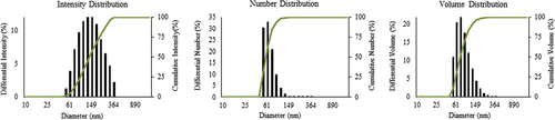
Figure 5. Antimicrobial activity of silver nanoparticles reaction mixture (100 μL), against S. aureus (a), S. enterica (b), V. parahemolyticus (c), C. albicans (d), and B. anthracis (e).
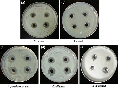
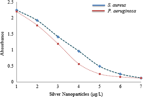
Figure 6. Biofilm inhibition activity of silver nanoparticles against S. aureus and P. aeruginosa.