Figures & data
Figure 1. (A) In the 24th week, the samples of the TEC group showed a smooth surface; the appearance was similar to the surrounding cartilage tissue, and the repaired tissue was well integrated with the surrounding normal cartilage. (B) In the 24th week, the defect site of Nano-HA/PLLA group was repaired basically, but the surface was uneven, showing an obvious boundary with the surrounding tissue. (C) In the 24th week, the defect of the control group became smaller, but there was still a pit in the center, showing an obvious boundary with the surrounding tissue.
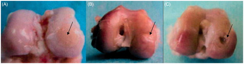
Figure 2. (A) In the 24th week, the chondrocytes stained by HE were increased in the TEC group. The matrix increased, and the pores of the scaffold were filled, but there was fissure on one side of the integrated area, with an obvious tidal line (×40). (B) In the 24th week, the cells stained by HE in the Nano-HA/PLLA group were decreased, with poor cellular layer. The surface tissue was flimsy, with no matrix of chondrocytes. The pores of the scaffold material were visible, with no tidal line (×40). (C) In the 24th week, HE staining of the control group showed that the repaired tissues were mainly threadlike fibers. The surface was uneven, and there was still a pit in the center, with no secretion of cartilage matrix (×40).
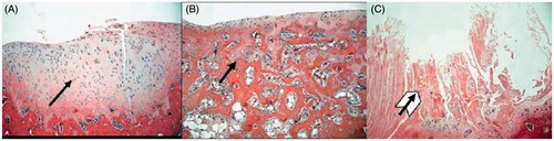
Figure 3. (A) In the 24th week, Safranine O staining of TEC group indicated the increase of matrix secreted by chondrocytes, with the matrix showing red color and the subchondral bone the green color. There were slight fissures on one side (×40). (B) In the 24th week, Safranine O staining of the Nano-HA/PLLA group showed that the surface tissue was thin and dominated by fibrocytes. The pores of the scaffold were visible, with no matrix secreted by the chondrocytes (×40). (C) In the 24th week, Safranine O staining of the control group indicated that the surface tissue was mainly composed of threadlike fibers and sunken in the center, with no matrix of chondrocytes (×40).
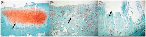
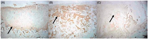
Figure 4. (A) In the 24th week, the type-II collagen immunohistochemical staining of the TEC group showed strongly positive result, and the scope of staining was consistent with that of HE staining in the 24th week (×40). (B) In the 24th week, the type-II collagen immunohistochemical staining of the Nano-HA/PLLA group showed that the pores of scaffold were still visible (×40). (C) In the 24th week, the type-II collagen immunohistochemical staining of the control group showed that the center was sunken and the margins were uneven (×40).