Figures & data
Table 1. Primer sequences used in PCR.
Figure 1. Characteristics of human ADSCs in vitro. (A) Proliferation potential of human ADSCs under the cultural conditions in vitro. (B) Population doubling time (PDT) of human ADSCs, the result showed that PDT was no significant change among seven consecutive culture passages.
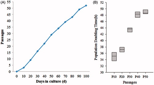
Figure 2. Special gene characteristics of human ADSCs. Immunoflourescence staining results showed that human ADSCs were positive for the CD13, CD71, CD73, CD90, CD105, and CD166, but negative for liver-associated genes, ALB and CYP3A4 (Scan bar = 50 μm).
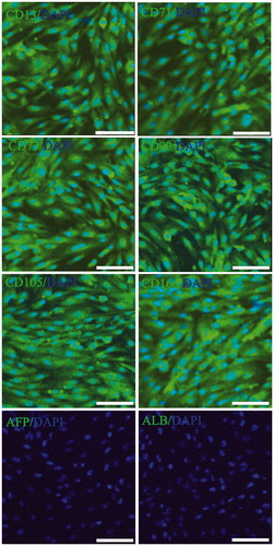
Figure 3. Special gene expression in different passages human ADSCs. CD13, CD71, CD73, CD90, CD105, and CD166, the special genes of ADSCs were analyzed using real time PCR in P0, P10, P20, P30, and P50, the result showed that these genes ware no significant change among seven consecutive culture passages.
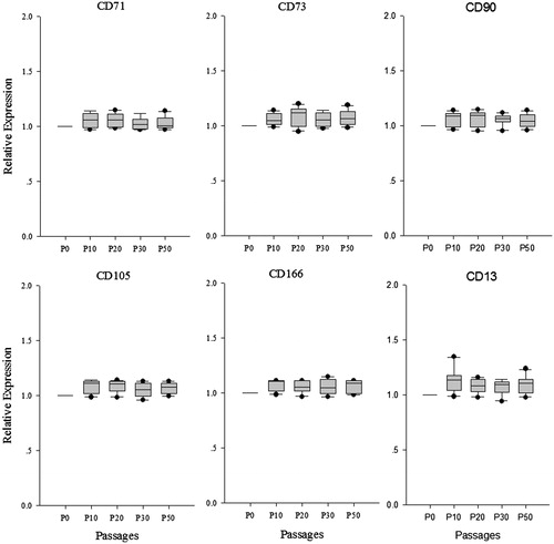
Figure 4. Identification surface markers of hepatocyte using immunoflourescence staining. Hepatocytes are the chief functional cells of the liver and perform an astonishing number of metabolic, endocrine, and secretory functions. ALB functions primarily as a carrier protein for steroids, fatty acids, and thyroid hormones and plays a role in stabilizing extracellular fluid volume. CYP3A4 is an important enzyme in the body, mainly found in the liver and in the intestine, which is a member of the cytochrome P450 superfamily of enzymes. The cytochrome P450 proteins are monooxygenases that catalyze many reactions involved in drug metabolism and synthesis of cholesterol, steroids, and other lipids components. Immunoflourescence staining result showed that ALB and CYP3A4 ware double positive in hepatocytes derived from ADSCs.
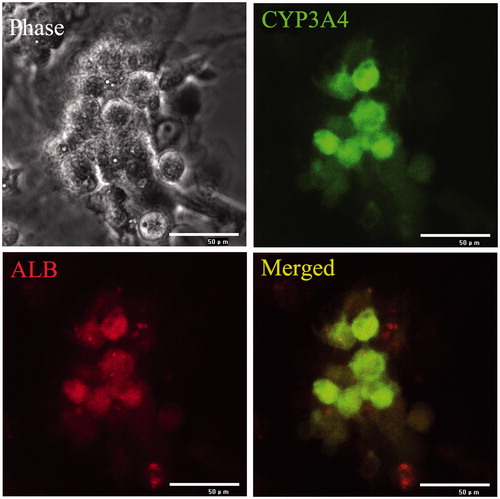
Figure 5. Gene expression of liver-associated genes detected in continuously cultured human ADSCs. Gene expressions in each time point were determined by real-time PCR and then normalized to that of control (0 day). The result showed relative expression level liver-associated genes mRNA were higher in differentiated hepatocyte compared to untreated control from day 14 of culture. At day 20, these genes mRNA levels were significantly increased (P < 0.05) in differentiated hepatocyte compared to control.
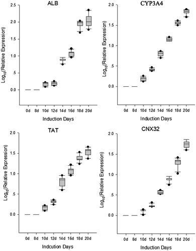
Figure 6. Transplantation of hADSCs, hADSCs-derived hepatocyte resulted in mouse acute liver injury. To test whether transplantation of hepatocyte and hADSCs taken part in recovery of hepatic lobule, paraffin section was used to observed the location for transplantation of hepatocyte in scathing liver tissue. Human nuclei protein was used as a label to discover the hADSCs or hADSCs-derived hepatocyte in tissue of hepatic lobule. The results showed that the human cells were found in mouse liver tissue through IV, but the group of hADSCs was significantly higher than group of hADSCs-derived hepatocyte. (A) Hematoxylin and eosin staining of paraffin section in acute liver injury. (B, C, and D, long arrow: central veins, short arrow: apoptotic hepatocytes) Representative immunofluorescence images of liver 3 weeks post-transplantation, stained for human nuclei protein to confirm the presence of hADSCs. Arrow: central veins. (bar = 100 μm).
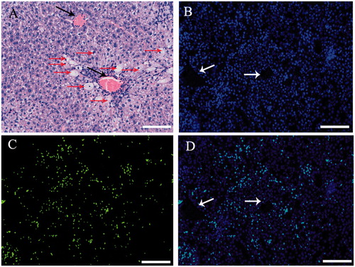
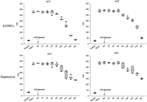
Figure 7. AST and ALT detection after injury and transplantation. ALT and AST were significantly increase after acute liver injury through serological testing by ELISA, the concentrate of ALT and AST were significantly decrease after injected ADSCs 8 days, but the hepatocyte group were observed changes after 12 days. Three weeks later, the concentrate of ALT and AST of ADSCs-treated mice reduced to a normal level (<50 U/L). In contrast, the ALT and AST concentrate of the injected hepatocyte mice was still above 100 U/L, and these mice could survive for nearly 4 weeks. These results showed that the ADSCs or ADSCs-derived hepatocyte, when injected into the vein, could improve liver function repair and functionally rescue the CCL4-treated mice with liver injury, but the ADSCs transplantation was better than ADSCs-derived hepatocyte transplantation.