Figures & data
Figure 1. Architecture of electrospun PCL nanofiber scaffold as seen under a scanning electron microscope at 4.00K×.
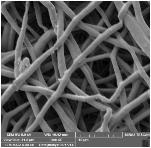
Figure 2. Morphology of IPFP-ASCs in culture as observed with an inverted phase-contrast microscope at passage2, 10×.
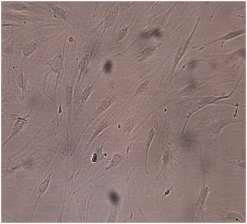
Figure 3. A photograph showing PCM in chondron as a bluish surrounding. Stained with toluidine blue, 40×.
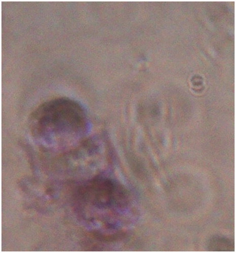
Figure 4. IPFP-ASCs attached to the PCL scaffold as seen by scanning electron microscope, 1.54K×.
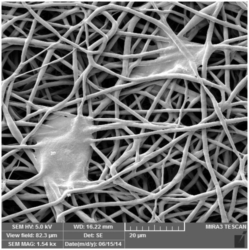
Figure 5. Cells were tested against human antigens CD31, CD44, CD45, CD90. All experiments were conducted at passage 2.
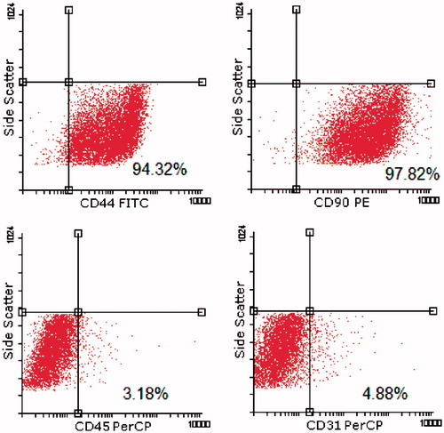
Figure 6. Relative gene expression of chondrogenic cell/PCL and cell/PCL coculture. Coculture conditions decreased expression of collagen2a1, aggrecan and increased expression of IHH. * = P < 0.05.
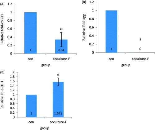
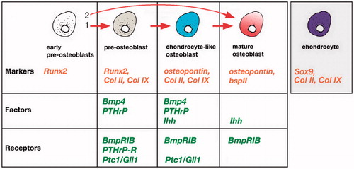
Figure 7. Regulation of osteoblastic differentiation in cranial dermal bone. Chondrocyte-like osteoblast (CLO) cells expressing a unique combination of collagen 2,9 (but not Sox9 or aggrecan) as well as Indian hedgehog (Ihh), Ptc1, Gli1, Bone morphogenic protein4 (Bmp4) and parathyroid hormone-related protein (PTHrP). As these cells develop into mature osteoblasts, they downregulate collagen 2, 9 expression.