
Figures & data
Figure 1. Experimental rapid prototype (RP) models. (A) Mandibular RP model. (B) Fibula RP model.
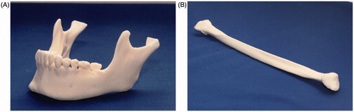
Figure 2. Mandibular reconstruction simulation and 3-D printed model. (A) The mandible was reconstructed with 3D imaging; the mandible was cut from the left subcondylar neck region to the right first premolar anterior region. Three-dimensional simulation of the mandible reconstruction with the fibula bony segments required bending the fibula at the canine area and at the angle area. This required three separate fibula segments: the anterior (light orange), the middle (brown), and the posterior (yellow) segments. (B) The reconstructed mandibular model was produced with a 3D printer, based on data from the simulated mandibular reconstruction with the fibula.
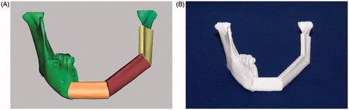
Figure 3. Fibula cutting guide template. (A) The fibula cutting guide template was prepared, based on the computer-aided design of a mandibular reconstruction with the fibula. The template (dark blue) includes sleeves that wrap around the fibula, and small sheets (upright squares) that indicate the cutting angles. (B) The fibula cutting guide template was fixed to the intact fibula (RP model). The cutting guide template was prepared with a 3D printer and biomaterials.
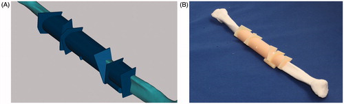
Figure 4. Experimental reconstruction of the mandibular RP model with fibula segments. The segments were cut with the manual method or with the STL fibula cutting guide. The metal miniplates are fixed to each joint.
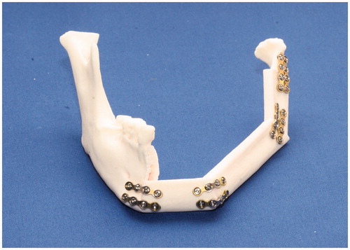
Figure 5. The right mandibular regions were registered to compare data from two images. A comparison is shown between the surgical simulation (red) result and the postoperative result (green).
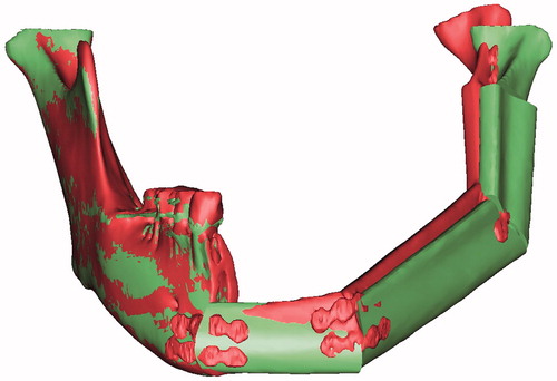
Figure 6. Reference planes for measuring distances. The axes (e.g. blue lines; model, red dotted line; simulation in the middle segment) drawn through the shafts of the fibula fragments were set to measure the angular errors (e.g. yellow fan in the middle segment) in the fibula bone segments. Vertical distance errors were evaluated at the measurement points, with reference to the FH plane (e.g. black dotted arrow line; the vertical distance from the medial point on the middle segment). Anteroposterior distance errors were evaluated at the measurement points with reference to the coronal plane. Lateral distance errors were evaluated at the measurement points, with reference to the sagittal plane. The errors were determined by comparing the preoperative surgical simulation results to the postoperative results.
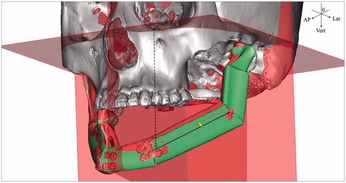
Table 1. Differences in 3-D distances (mm) between the simulated (planned) surgical result and two models created with different fibula cutting methods for mandibular reconstruction.
Table 2. Axis angles (°) of the fibula segments between the simulated (planned) surgical result and two models created with different fibula cutting methods for mandibular reconstruction.
Table 3. Differences in vertical distances (mm) between the simulated (planned) surgical result and two models created with different fibula cutting methods for mandibular reconstruction.
Table 4. Differences in anteroposterior distances (mm) between the simulated (planned) surgical result and two models created with different fibula cutting methods for mandibular reconstruction.
Table 5. Differences in lateral distances (mm) between the simulated (planned) surgical result and two models created with different fibula cutting methods for mandibular reconstruction.