
Figures & data
Table 1. Patient characteristics.
Figure 1. Intraoperative navigation view of Case 4. The embolization coil is a bright point at the intersection of the red lines, indicated by the yellow circle. It was easily palpable and helpful as a reference point.
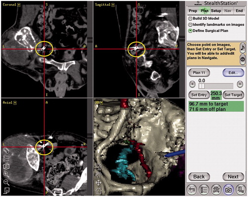
Figure 2. Intraoperative navigation view and postoperative CT 34 months after operation (Case 1). The inferior gluteal artery is indicated at the outlet of the greater sciatic notch with a dark blue line (a). The inferior gluteal artery (yellow arrows) on the operated side was preserved (b, c).
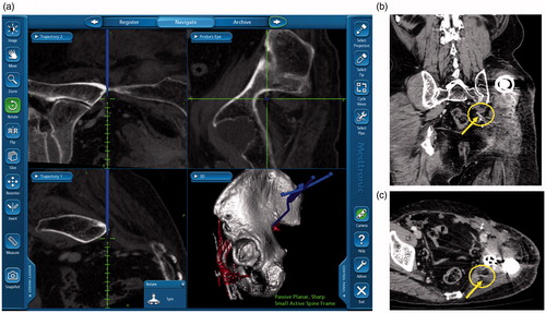
Figure 3. Intraoperative navigation view and postoperative CT 12 months after operation (Case 4). The inferior gluteal artery was detected on the pelvic cavity side of the greater sciatic notch (a). The patent inferior gluteal artery on the operated side (b, c).
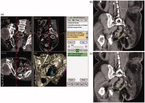
Figure 4. Intraoperative navigation view of Case 2. Arrowhead indicates S2 root and arrow indicates S1 root encased by tumor.
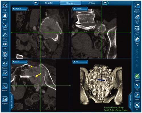
Table 2. Outcomes.