6,546
Views
36
CrossRef citations to date
0
Altmetric
Clinical Research

Diagnosis and treatment of sleep disorders: a brief review for clinicians

Diagnóstico y tratamiento de los trastornos del sueño: una breve revisión para los clínicos

Diagnostic et traitement des troubles du sommeil: brève revue pour les cliniciens

&
Pages 371-388 | Published online: 01 Apr 2022

Figures & data

Table I Classification of sleep disordersCitation4. NOS, not otherwise specified; REM, rapid eye movement.

Table II Multicomponent therapy instructions

Table III Chronotherapy instructions to advance sleep phase.Citation47

Figure 1. Hypopnea in a patient with obstructive sleep apnea syndrome. Note the low amplitude signals seen in the nasal cannula and airflow channels with increasing effort demonstrated on the chest and abdominal (Abd) channels. The Pes (esophageal pressure [PES]) channel shows crescendo increases in esophageal pressure with reversal.

Table IV Clinical features of obstructive sleep apnea syndrome.
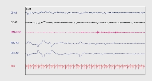
Figure 2. Sleep-onset rapid eye movement (REM) during an mean sleep latency test (MSLT) nap in a patient with narcolepsy. Electroencephalogram (EEG) leads (C3-A2 and 02-A1) demonstrate low voltage mixed frequency theta activity. EMG-Chin shows atonia with phasic events. Electrooculography (EOG) demonstrates REM.