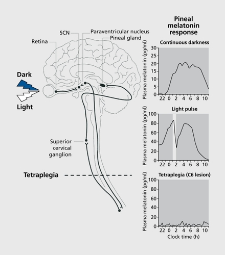
2,291
Views
129
CrossRef citations to date
0
Altmetric
Clinical Research

Visual impairment and circadiam rhythm disorders

Deterioro visual y transtornos de los ritmos circadianos

Troubles du rythme circadien et déficit visuel

, &
Pages 301-314 | Published online: 01 Apr 2022

Figures & data

Figure 1. Neuroanatomy of the circadian system. Light is detected by specialized retinal photoreceptors and transduced to the circadian pacemaker in the hypothalamic suprachiasmatic nuclei (SCN) via the monosynaptic retinohypothalamic tract (RHT). SCN efferents project to the pineal gland via the paraventricular nucleus (PVN) and the superior cervical ganglion (SCG). Under normal light-dark conditions, the pineal melatonin rhythm peaks at night during the dark phase with the duration of melatonin production reflecting the scotoperiod (night-duration) (top panel). When the eyes are exposed to light at night however, melatonin production is immediately suppressed, recovering once the light is switched off (middle panel). The “melatonin suppression test” confirms the functional integrity of the retina-RHT-SCN-PVN-SCG-pineal pathway. In individuals in whom the upper spinal cord has been severed, for example in tetraplegie patients, the signal from the SCN cannot reach the pineal gland and no rhythmic production of melatonin is possible (lower panel). Circadian rhythms in other parameters that do not require the SCN-SCG pathway, such as Cortisol and TSH rhythms, remain fully intact in tétraplégie patients.Citation1,Citation2 Adapted from reference 3: Wehr TA, Duncan WC Jr, Sher L, et al. A circadian signal of change of season in patients with seasonal affective disorder. Arch Gen Psychiatry 2001;58:1108-1114. Copyright © American Medical Association 2001; and from reference 4: Zeitzer JM, Ayas NT, Shea SA, Brown R, Czeisler CA Absence of detectable melatonin and preservation of cortisol and thyrotropin rhythms in tetraplegia. J Clin Endocrinol Metab. 2000;85:2189-2196. Copyright © Endocrine Society 2000
Figure 2. Characterizing circadian phase using urinary aMT6s rhythms. Examples of urinary aMT6s rhythms measured for 48 hours over 4 consecutive weeks are shown for three representative blind subjects. Panel A shows a normally entrained aMT6s rhythm with a normal night-time peak (grey bar, normal range 1:00 - 7:00 h), that remains remarkably stable from week to week, despite the study being conducted by the subject at home. A proportion of subjects exhibit stable 24-hour rhythms but are entrained to an abnormally advanced or delayed phase. Panel B shows an advanced subject who has an aMT6s rhythm peaking relatively early compared with normal. Panel C shows the most common rhythm type observed in subjects with no conscious perception of light (NPL), namely a nonentrained “free-running” rhythm. While the rhythm may initially appear arrhythmic, closer inspection shows that the aMT6s peak is changing from week to week over the 4-week study (W1 - W4), getting relatively later each week, and thereby exhibiting a circadian period >24 hours (τ=24.7 h in this case). In these cases, longitudinal assessment over several weeks or even months, if the endogenous period is very close to 24 hours, is required to distinguish the nonentrained rhythm from a normally or abnormally entrained stable rhythm. Adapted from reference 64: Skene DJ, Lockley SW, James K, Arendt J. Correlation between urinary Cortisol and 6-sulphatoxymelatonin rhythms in field studies of blind subjects. Clin Endocrinol. 1999; 50: 715-719. Copyright © Blackwell Publishing 1999
Figure 3. Circadian rhythms of sleep and melatonin in two blind subjects. The left-hand panels show subjective sleep times (■) over 4 to 5 weeks, double-plotted according to time of day (abscissa) and study day (ordinate). Subjects also collected sequential 4- to 8-hourly urine samples for 48 hours per week and the peak time of urinary 6-sulphatoxymelatonin (aMT6s) production each week (○) is plotted. The right-hand panels show the weekly rhythms of urinary aMT6s production (●) that correspond to the peak times shown and the gray bars represent the normal range of peak production for aMT6s (1:00 - 7:00 h). Panel A shows data from a visually impaired female aged 35 years with a visual acuity allowing her to detect hand movements only and normal circadian rhythms (Subject 21). Both the sleep-wake cycle and aMT6s rhythm remain entrained to 24 hours (A, left panel) with normal night-time production of melatonin (A, right panel) and such rhythms would be typical of most sighted subjects. Note that her visual impairment while severely disrupting the visual system, does not affect the circadian photoreception system, illustrating the functional separation of the visual and circadian photoreceptor systems. Panel B shows the rhythms from a 66-year-old totally blind man with one eye and no conscious light perception (Subject 20). He exhibits a sleep-wake cycle and melatonin rhythm that is not entrained to 24 hours. The aMT6s rhythm exhibits a non-24-hour period (τ = 24.68 h) which clearly delays from week-to-week and is not entrained to the night (B, right panel). Similarly the sleep pattern show a cyclic sleep disorder characteristic of “non-24-hour sleep-wake disorder,” with recurrent episodes of disturbed night-time sleep and many daytime naps when melatonin peaks during the day (eg, Days 1 to 8, or 28 to 35) that alternate with episodes of good sleep and high day-time alertness when the melatonin is produced during the night (eg, Days 10-25) (B, left panel). Reproduced from reference 68: Lockley SW. Human circadian rhythms: influence of light on circadian rhythmicity in humans. In: Squire LR, ed New Encyclopaedia of Neuroscience, Oxford, UK: Elsevier. In press. Copyright © Elsevier 2007.
Figure 4. Relationship between sleep timing and circadian phase in entrained blind subjects. Panel A shows subjective sleep (■) and urinary 6-sulphatoxymelatonin rhythm (○) timing over 4 weeks (study day on ordinate axis, clock time on abscissa) in two blind subjects, one with advanced sleep phase syndrome (ASPS) and one with delayed sleep phase syndrome (DSPS). In the ASPS subject the advanced aMT6s peak (~ 20:00 h, 8 hours earlier than normal) is associated with many daytime naps in the late afternoon and early evening, as the circadian system attempts to initiate sleep during the biological night, and an early wake-time from the main sleep episode. The DSPS subject exhibits a relatively delayed sleep onset time on most days, but shows a particularly delayed sleep pattern during weekends when not having to set an alarm for work, as on other days. Panel B shows the correlation between the timing of the aMT6s peak (abscissa) and sleep onset time (ordinate) in visually impaired entrained subjects, illustrating the change in sleep timing associated with altered circadian phase. Adapted from reference 62: Lockley SW, Skene DJ, Butler U, Arendt J. Sleep and activity rhythms are related to circadian phase in the blind. Sleep. 1999;22:616-623. Copyright © Associated Professional Sleep Societies 1999.
Figure 5. Absence and presence of circadian photoreception in two totally blind subjects. Panels A and C: Subjects completed daily sleep and nap diaries for ~11 to 12 weeks and their sleep times (solid lines) are double-plotted according to convention in Figure 3. Subjects also had their circadian phase measured at regular intervals from either core body temperature minimum ((+) Panel A) or plasma melatonin onset (Δ Panel C). Panels B and D show the results of a melatonin suppression test during which the subjects' eyes were exposed to bright white light (6000 to 13 700 lux) for 90 to 100 minutes during plasma melatonin production. Panels A and B show the sleep-wake pattern and melatonin suppression response for a 70-year-old blind man with congenital glaucoma. He retained both eyes but reported no conscious light perception. Electroretinogram (ERG) and visually evoked potential (VEP) responses were not detectable. As shown in Panel A, the subject's circadian system was not entrained to the light-dark cycle and the core body temperature rhythm exhibited a non-24-hour period. Consistent with the lack of entrainment by light light exposure did not have any effect on the melatonin rhythm, confirming that the retina-SCN-pineal pathway was not functional in this patient. Panels C and D show the sleep-wake pattern and melatonin suppression results for a 21-year-old woman with Leber's congenital amaurosis, a type of retinal dystrophy The ERG was undetectable but an abnormal VEP was recorded. As shown in Panel C, the sleep-wake cycle was normal with stable entrainment to 24 hours. The melatonin suppression response was clearly positive, with an immediate suppression of melatonin at lights on (open bar) which ceased once the bright lights were switched off. Despite the subject's lack of conscious light perception, light was still able to stimulate the retina-SCN-pineal pathway; covering the eyes prevented the response. The normal sleep-wake cycle, coupled with the positive melatonin suppression test indicated that light was able to entrain the circadian system of this totally blind woman. Note: a similar sleep-wake pattern but accompanied by a negative melatonin suppression test would indicate that (i) the subject was entrained to 24 hours by a nonphotic time cues or (ii) the subject's endogenous circadian period was exactly 24 hours. SCN, suprachiasmatic nucleus. Reproduced from reference 74: Czeisler CA, Shanahan TL, Klerman E., et al. Suppression of melatonin secretion in some blind patients by exposure to bright light. N Engl J Med. 1995;332:6-11. Copyright © Massachusetts Medical Society 1995
Figure 6. Entrainment of circadian rhythms in the blind with melatonin. This Figure shows the double-plotted sleep timing (■) and urinary Cortisol peak times (○) for two totally blind men treated with 5 mg melatonin PO at 21:00 h for at least one circadian cycle in a placebo-controlled single-blind design. Sequential study days are plotted on the ordinate and clock time is plotted on the abscissa. The start and end of placebo (Pla) and melatonin (Mel) treatment are indicated by the boxes. Panel A shows a subject who was entrained by melatonin treatment (S17). He exhibited a cortisol period of 24.3 h during placebo treatment but was fully entrained (τ=24.0 h) at a normal circadian phase (mean ± SD Cortisol peak time =9.9±0.7 h)Citation67 during melatonin administration. The stabilized sleep-wake cycle observed during melatonin treatment becomes immediately disrupted upon cessation of treatment and reverts to the characteristic non-24-hour pattern, with cyclic episodes of good sleep at night and a small number of daytime naps followed by extremely disrupted night-time sleep and excessive day-time napping. Panel B shows a subject who failed to entrain to melatonin treatment (S45) and who exhibited similar periods during melatonin (25.2 h) and placebo (24.9 h) administration, despite melatonin treatment for nearly two full circadian cycles. The persistent cyclic non-24-hour sleep disorder is apparent throughout both the placebo and melatonin treatment. Reproduced in part from ref 109: Lockley SW, Skene DJ, James K, Thapan K, Wright J, Arendt J. Melatonin administration can entrain the free running circadian system of blind subjects. J Endocrinol. 2000; 164,:R1-R6. Copyright © Society for Endocrinology 2000