Marc ThiouxNetherlands Institute for Neuroscience, The Netherlands Royal Academy of Science (Koninklijke Nederlandse Akademie van Wetenschappen), The NetherlandsCorrespondence[email protected]
&
Christian KeysersNetherlands Institute for Neuroscience, The Netherlands Royal Academy of Science (Koninklijke Nederlandse Akademie van Wetenschappen), The Netherlands
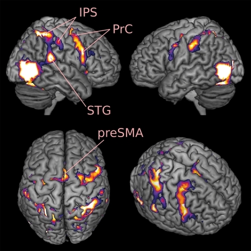
Figure 1. Parietal and premotor cortices are active during the observation of hand actions. IPS, Intraparietal sulcus; PrC, Precentral gyrus/sulcus; preSMA, pre-supplemetary motor area; STG, superior temporal gyrus. Results are from a random effect analysis of the functional images of 17 participants (P<0.0005 FWE corrected; F3,48 = 20.34). The most anterior part of the IPS cluster extends to Brodmann area 2 (in the anterior IPS aka post-central sulcus), the highest level of processing in the primary somato-sensory cortex.
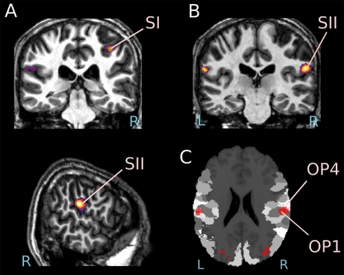
Figure 2. Activation of the primary and secondary somatosensory cortices in a single subject observing someone touching an object Functional images are superimposed on the subject's own anatomy (P<0.05 FDR corrected). (A) Coronal slice 33 mm posterior to the anterior commissure showing the activation of Brodmann area 2 of the primary somatosensory cortex (SI). (B) Coronal plane 26 mm posterior to the anterior commissure showing the activation of the secondary somato-sensory cortex (SII), and sagittal view of the right hemisphere at 60 mm from the midline showing the same cluster. (C) Overlay of SI activity on an anatomical probability map showing the location of OP1 and OP2 (SPM Anatomy Toolbox)
Table I. Six FMRI studies investigating face processing in participants with autism spectrum disorders (ASD) and typically developing (TD) individuals, and providing whole brain results. ASD, participants with autism spectrum disorder; TD, typically developing individuals; BA44/45, inferior frontal gyrus pars opercularis/triangularis; PrC, Precentral gyrus/sulcus; MPFC, medial prefrontal cortex; OFC, orbito-frontal cortex; vPMC, ventral premotor cortex; PoC, post-central gyrus/sulcus; STS, superior temporal sulcus; IFG, inferior frontal gyrus; *, Talairach coordinates
DaprettoM.DaviesMS.PfeiferJH.et al.Understanding emotions in others: mirror neuron dysfunction in children with autism spectrum disorders.Nat Neurosci.20069283016327784
BookheimerSY.WangAT.ScottA.SigmanM.DaprettoM.Frontal contributions to face processing differences in autism: evidence from fMRI of inverted face processing.J Int Neuropsychol Soc.20081492293218954473
UddinLQ.DaviesMS.ScottAA.et al.Neural basis of self and other representation in autism: an FMRI study of self-face recognition.PLoS ONE.20083e352618958161
AshwinC.Baron-CohenS.WheelwrightS.O'RiordanM.BullmoreET.Differential activation of the amygdala and the 'social brain' during fearful face-processing in Asperger Syndrome.Neuropsychologia.20074521416806312
PierceK.HaistF.SedaghatF.CourchesneE.The brain response to personally familiar faces in autism: findings of fusiform activity and beyond.Brain.2004127(Pt 12)2703271615319275