909
Views
39
CrossRef citations to date
0
Altmetric
Clinical Research

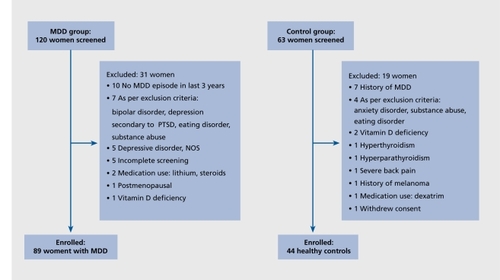
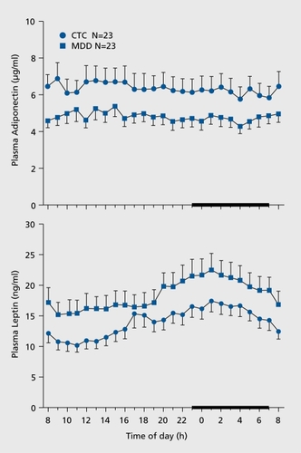
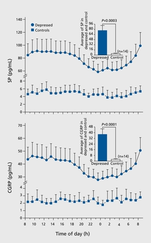
Major depressive disorder is a risk factor for low bone mass, central obesity, and other medical conditions

El trastorno depresivo mayor es un factor de riesgo para una masa ósea disminuida, obesidad central y otras condiciones médicas

Le trouble dépressif majeur est un facteur de risque de faible masse osseuse, d'obésité centrale et d'autres pathologies

Pages 73-87 | Published online: 01 Apr 2022

Figures & data

Figure 1. Study flow diagram. The number of subjects screened, reasons for exclusion, and number of subjects enrolled in the study are listed. Reproduced from ref 5: Eskandari F, Martinez PE, Torvik S, et al. Premenopausal, Osteoporosis Women, Alendronate, Depression (POWER) Study Group Low bone mass in premenopausal women with depression. Arch Intern Med. 2007;167:2329-2336. Copyright © American Medical Association 2007
Figure 2. Meta-analysis forest plots for AP spine bone mineral density (BMD), total femur BMD and femoral neck BMD. (A) AP spine BMD: Most studies show lower AP spine BMD in depressed subjects, with 3 studies indicating that BMD was higher in controls. (B) Total femur: Most studies show lower total femur BMD in depressed subjects, with 3 studies indicating that BMD was higher in controls, and 2 studies showing no difference. (C) Femoral neck BMD: Most studies show lower femoral neck BMD in depressed subjects, with three studies indicating that BMD was higher in controls. For each one of these three graphs under the first column are listed in chronological order the studies included and the year of publication; these studies are subdivided in studies conducted only in women, only in men, and studies conducted in men and women. Studies that utilized DSM criteria to characterize depression are indicated with a dagger. In the last column the relative weight of each individual study is reported. Reproduced from ref 12: Cizza G, Primma S, Coyle M, Gourgiotis L, Csako G. Depression and osteoporosis: a research synthesis with meta-analysis. Horm Metab Res. 2010;42:467-482. Copyright ©Thieme 2010
Figure 3. Plasminogen activator-1 (PAI1). PAI-1 concentrations exhibit an exponential distribution both in subjects with MDD and controls. Panel A: 0800 h PAI-1 concentration. Panel B: 2000 h PAI-1 concentration. Reproduced from ref 29: Eskandari F, Mistry S, Martinez PE, et al; POWER (Premenopausal, Osteopenia/Osteoporosis, Women, Alendronate, Depression) Study Group. Younger, premenopausa women with major depressive disorder have more abdominal fat and increased serum levels of prothrombotic factors: implications for greater cardiovascular risk. Metabolism. 2005;54:918-924. Copyright © W. B. Saunders 2005
Figure 4. Analysis of baseline C-reactive protein (CRP) levels in women with major depressive disorder (MDD) and healthy control women Upper panel: Box plots showing median, quartiles, and extreme values. The box represents the values between the 25th and 75th percentiles. The horizontal bar across each box represents the median value. Asterisks represent extreme values (values more than 3 box lengths from the upper edge of box). Open circles represent outliers (values between 1.5 and 3 box lengths from upper edge of box). One depressed subject with high CRP levels (29.3 mg/L) had reported recovery from an acute infection at the time of visit and was therefore excluded from the analyses. Lower panel: Percentile distribution of CRP values. CRP values for select percentiles are shown in the inset table. At the uppermost percentile (75th), women with MDD have CRP levels over twice as high as control women. The dashed line marks the CRP level of 10 mg/L above which there are only MDD subjects. Reproduced from ref 35: Cizza G, Eskandari F, Coyle M, et al; P.O.W.E.R (Premenopausal, Osteoporosis Women, Alendronate, Depression) Study Group. Plasma CRP levels in premenopausal women with major depression: a 12-month controlled study. Horrn Metab Res. 2009;41:641-648 Copyright © Thieme 2009
Figure 5. Mean 24-hour plasma adiponectin and leptin concentrations in women with major depressive disorder (MDD, N=23) and Controls (N=23). Upper panel adiponectin concentrations: In both groups, adiponectin exhibited a circadian variation characterized by slightly higher values during early afternoon with a peak around 1400 h. Adiponectin remained lower in women with MDD over 24 h. 0800 h and 24 h mean adiponectin was significantly lower in women with MDD compared with controls (0800 h P adjusted for weight =0.0053; mean 24-h P adjusted for weight =0.042; analysis of covariance). Error bars represent SEM. Lower panel leptin levels. In both groups, leptin exhibited a circadian variation characterized by a nocturnal zenith around 0200 h and a nadir around 1 000 h. Leptin remained higher in women with MDD over 24 h 0800 h and 24 h mean leptin was significantly higher in women with MDD compared with controls (0800 h P adjusted for weight =0.010; mean 24-h P adjusted for weight =0.021; analysis of covariance). Mean nocturnal (2000 h-0400 h) leptin, was greater in women with MDD than controls (21.05 + 0.91 ng/mL vs 16.10 + 0.98 ng/mL, respectively, P<0.001). Error bars represent SEM Reproduced from ref 47: Cizza G, Nguyen VT, Eskandari F, et al; POWER Study Group. Low 24-hour adiponectin and high nocturnal leptin concentrations in a case-control study of community-dwelling premenopausal women with major depressive disorder: the Premenopausal, Osteopenia/Osteoporosis, Women, Alendronate, Depression (POWER) study. J Clin Psychiatry 2010;71:1079-1087. Copyright © Physicians Postgraduate Press 2010
Figure 6. Plasma levels of substance P (SP) and calcitonin-gene-relatedpeptide (CGRP). Mean 24-h levels of SP (upper panel) and CGRP (lower panel) were lower in women with depression compared with controls. Reproduced from ref 39: Hartman JM, Berger A, Baker K, et al. Quality of life and pain in premenopausal women with major depressive disorder: The POWER Study. Health Qual Life Outcomes. 2006;4:2. Copyright © Biomed Central 2006