5,123
Views
91
CrossRef citations to date
0
Altmetric
Translational Research

Neuroimaging in anxiety disorders

Neuroimágenes en los trastornos ansiosos

Neuro-imagerie dans les troubles anxieux

&

Figures & data

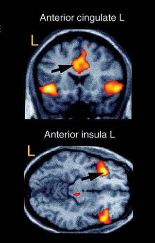
Figure 1. Activation in the left anterior cingulate cortex (top) and left anterior insula (bottom) during presentation of conditioned (vs neutral) faces. Reproduced from ref 5: Buchel C, Morris J, Dolan RJ, Friston KJ. Brain systems mediating aversive conditioning: an event-related fMRI study. Neuron. 1998;20:947-957. Copyright © Elsevier, 1998
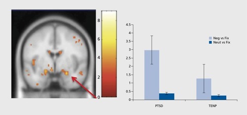
Figure 2. Activation in the right amygdala is enhanced in post-traumatic stress disorder (PTSD) patients compared with trauma-exposed non-PTSD participants (TENP) during the presentation of emotionally negative pictures. Fix, fixation baseline; Neg, negative; Neut, neutral. Reprinted from ref 43: Brohawn KH, Offringa R, Pfaff DL, Hughes KC, Shin LM. The neural correlates of emotional memory in posttraumatic stress disorder. Biol Psychiatry. 2010;68:1023-1030. Copyright © Elsevier, 2010
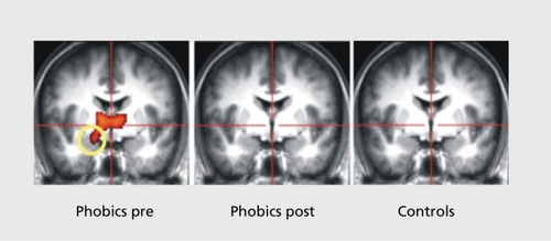
Figure 3. Amygdala activation during presentation of pictures of spiders (vs neutral pictures) in spider phobic subjects before and after successful treatment, and in non-phobic control subjects. Reprinted from ref 59: Goossens L, Sunaert S, Peeters R, Griez EJ, Schruers KR. Amygdala hyperfunction in phobic fear normalizes after exposure. Biol Psychiatry. 2007;62:1119-1125. Copyright © Elsevier, 2007