4,308
Views
60
CrossRef citations to date
0
Altmetric
Clinical Research

Neurofeedback and networks of depression

Retroalimentación neural y redes de la depresión

Neurofeedback et réseaux de la dépression

Figures & data

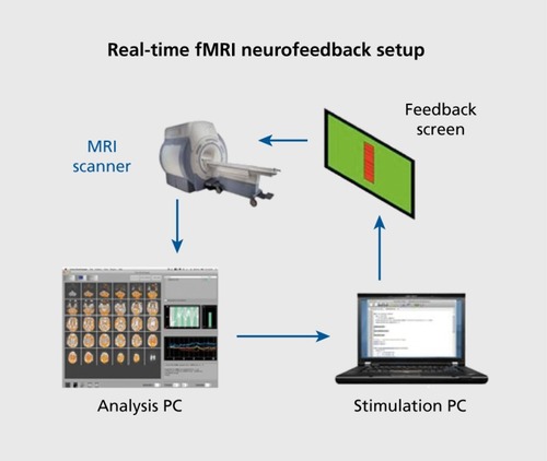
Figure 1. Basic diagram of a real-time functional magnetic resonance imaging brain-computer interface for neurofeedback. Figure courtesy of Isabelle Habes

Table I. Symptoms of depression. Five symptoms are required over a 2-week period for an episode of major depression (DSM-IV). ICD-10 defines depressive episodes by a combination of the most typical (printed in bold face) and other symptoms.Citation19 The number of symptoms determines the severity of the episode: 2 typical and 2 other: mild; 2 typical and 3 or 4 other: moderate; 3 typical and 4 or more other: severe. The column on the right indicates the broad domains into which the symptoms can be tentatively classified: ER, emotion regulation; C, cognition; M, motivation; H, homoeostasis
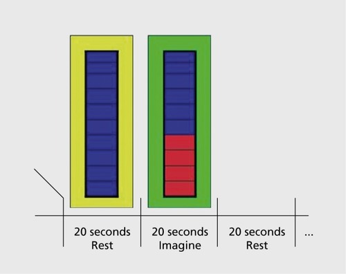
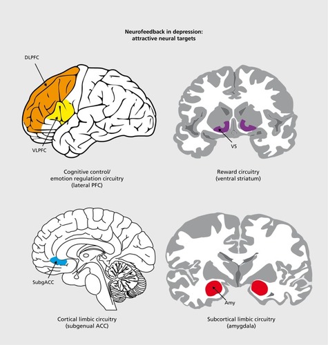
Figure 2. Display screen of visual neurofeedback with an outline of the protocol. The patients trained to increase activity in functionally localized areas during 20-second periods, alternating with 20 second periods of rest. Overall, they did this for 20 minutes each in four weekly sessions. Adapted from ref 43: Subramanian L, Hindle JV, Johnston S, et al. Realtime functional magnetic resonance imaging neurofeedback for treatment of Parkinson's disease. J Neuroscf. 2011:31:16309-16317. Copyright © Society for Neuroscience 2011
Figure 3. Cognitive-affective brain systems that could become targets for neuromoduiation in depression. DLPFC, dorsolateral prefronta! cortex; VLPFC, ventrolateral prefrontal cortex; ACC, anterior cinguiate cortex; Amy, amygdala Adapted from ref 38: Esmail S, Linden D. Cogn Sci. 2011 ;6. Copyright © Nova Science Publishers, Inc.