William G. Honera Departments of Psychiatry, University of British Columbia, Vancouver, British Columbia, CanadaCorrespondence[email protected]
,
Alfredo Ramos-Miguelb Department of Pharmacology, University of the Basque Country, and Centro de Investigación Biomédica en Red de Salud Mental, CIBERSAM, Madrid, Spain
,
Jehan Alamric Departments of Anaesthesia, Pharmacology & Therapeutics, University of British Columbia, Vancouver, British Columbia, Canada
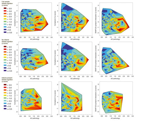
Figure 1. Global cognitive function nearest to death (z-score, controlled for age, sex, and educa- tion) in relation to levels of presynaptic proteins complexin-I and complexin-II, and SNAP-25-syntaxin protein-protein interaction, and to level of Alzheimer disease (AD) pathology. The top row illustrates the full sample (n= 420), middle row samples from brains free of infarcts (n=268), bottom row samples from brains with infarcts (n=152). SNAP-25, synaptosome-associated protein-25.
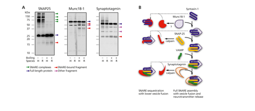
Figure 2. (A) Immunoblots of monomeric, full-length presynaptic proteins (blue arrows),
protein-protein complexes, and other fragments in denaturing gels. Boiling, rather than
PAGE denaturing chemicals, allows full SNARE complex dissociation. Fragments showing
greater immunoreactivity after SNARE disruption (boiled samples) are proposed to
participate in SNARE complex formation/modulation. (B) Complexes (right side of panel)
formed by full-length SNARE proteins (center of panel) and possibly sequestered into
other complexes (left side of panel) after enzymatic cleavage. PAGE, polyacrylamide gel
electrophoresis; SNAP-25, synaptosome-associated protein-25; Munc18-1, mammalian
unc-18-1; SNARE, soluble N-ethylmaleimide-sensitive factor attachment protein receptor;
VAMP, vesicle-associated membrane protein.