Figures & data
Fig. 1 Initial lower extremity radiographs showing the distal tibial and fibular fractures.
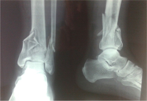
Fig. 2 Postoperative anteroposterior lower extremity radiograph showing the internal fixation of the pilon fracture.
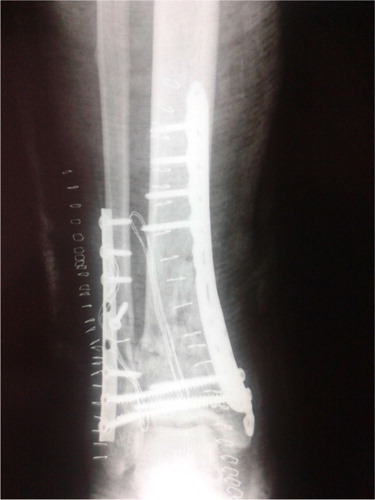
Fig. 3 Postoperative lateral lower extremity radiograph showing the internal fixation of the pilon fracture.
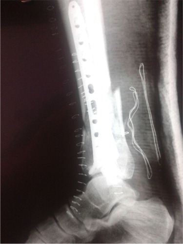
Fig. 4 Initial presentation of the skin lesion after the internal fixation of the pilon fracture.
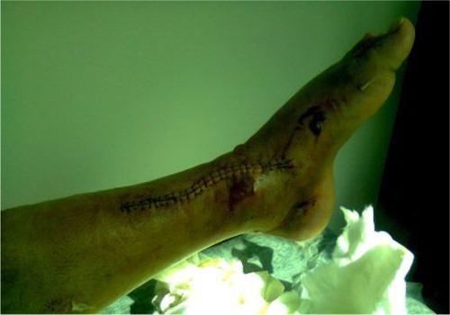
Fig. 5 Postoperative angiography showing partial obstruction of the fibular artery.
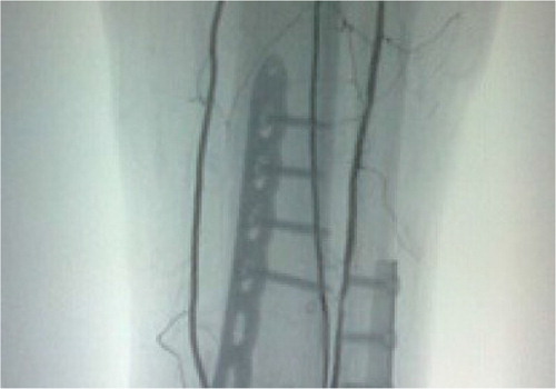
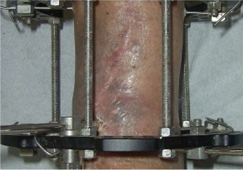
Fig. 6 Clinical picture showing the Ilizarov external fixation combined with Papineau technique.
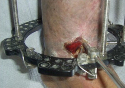
Fig. 7 Postoperative anteroposterior lower extremity radiograph at 6 months after the Ilizarov external fixation application.
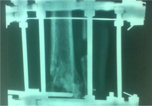
Fig. 8 Postoperative lateral lower extremity radiograph at 6 months after the Ilizarov external fixation application.
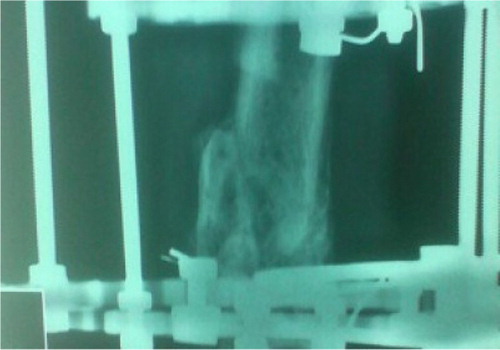
Fig. 9 Clinical picture showing the soft tissue defect healed at 6 months after the Papineau technique.