Figures & data
Fig. 1 Illustration of a life-threatening malignant central airway obstruction. CT images, 1–2: coronal and transversal slices showing almost complete occlusion of the right and left main bronchus by a tumour; video bronchoscopy, image 3: protruding tumour at the level of carina; CT images, 4–5: coronal and transversal slices showing the reestablished lumen of both main bronchi after stent implantation and radiotherapy.
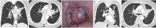
Fig. 2 The new removable stent (magnified) made by self-expanding nitinol with a thread that extends beyond the tubular part. The end of the thread can be placed at a distance and be used to unravel the device to the thread from which it was made and remove the stent.
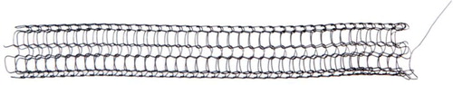
Fig. 3 The phantom model where the stent, introducer system and removability were tested before the animal study which took place at the Operating Room of the Future, ORF.
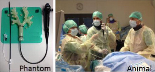
Table 1 Study design
Fig. 4 Acute study: Day 1 – placement and unravelling phase – placement of the stent, a (distant view) and b (close view). The unravelling procedure, midways and before complete removal, c and d, respectively.
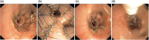
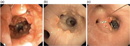
Fig. 5 Chronic study: (a) Day 14 – after removal of proximal stent, and polyps are seen, (b) Day 18 – same area 4 days later showing dramatic healing and (c) Day 18 – view of the residual distal stent.