Figures & data
Fig. 1 Immunohistochemical staining of histological biopsy from rectal mass: an adenocarcinoma positive for TTF-1 (A) and CK7 (B).
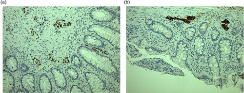
Fig. 2 Chest PET-CT scan showing no sign of a primary lung lesion but multiple enlarged lymph nodes, a left-sided compression atelectasis, and ipsilateral pleural effusion.
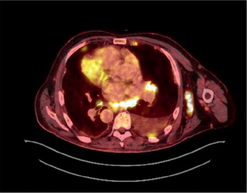
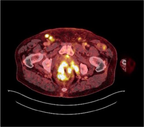
Fig. 3 Abdominal PET-CT scan showing a rectal mass, moderate quantities of ascites, and enlarged lymph nodes in pelvis and groin.