Figures & data
Fig. 1 (a) PA and (b) lateral views of the chest demonstrate a large left pleural collection with an air-fluid level (arrows). Small right pleural effusion is also seen.
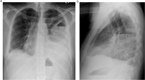
Fig. 2 CT scan of the chest showing the loculated left pleural fluid collection with air-fluid level (arrow) and the adjacent parenchymal consolidation.
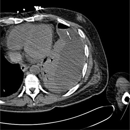
Fig. 3 Small bore chest tube was placed from an anterior approach.
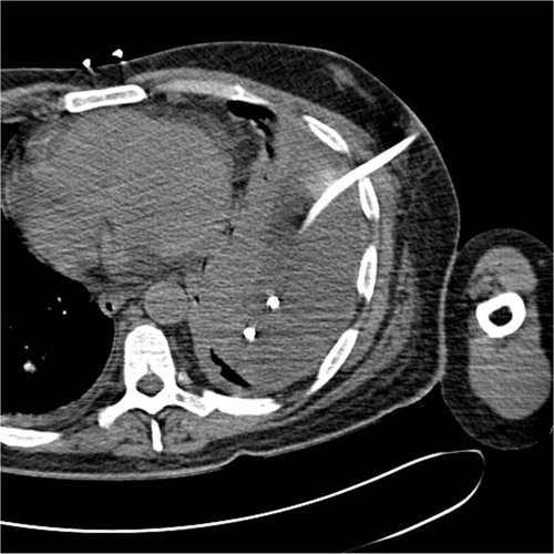
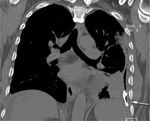
Fig. 4 Coronal image of the chest demonstrates a small bore chest tube partially surrounded by air in the left pleural space (arrow). No remaining fluid is seen. Adjacent lung consolidation has decreased.