Figures & data
Fig. 1 Computed tomography scan of the abdomen (axial view) showing thickening of the colon wall (short thick arrow, on the left) as well as right kidney mass (long thin arrow).
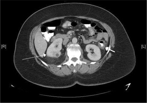
Fig. 2 Colonoscopic picture of splenic flexure showing edematous, erythematous, and friable mucosa.
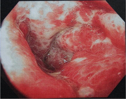
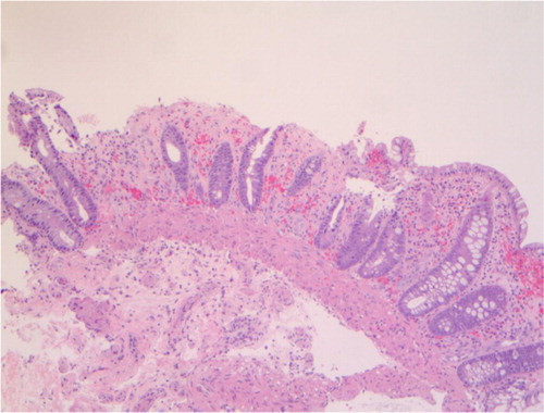
Fig. 3 Low-power photomicrograph (hematoxylin and eosin stain) of a biopsy of splenic flexure showing focal glandular dropout, small glands with mucin depletion, denudation of surface epithelium, stromal hemorrhage, and fibrosis.