Figures & data
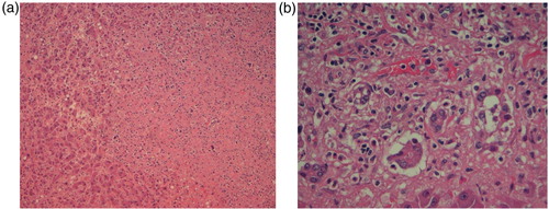
Fig. 1 (a) High power view of Level 7 lymph node from the neck showing atypical cells, some of which are multinucleated and consistent with Reed–Sternberg (RS) cells although not typical. (b) Immunohistochemical stain showing RS cells positive for CD30.
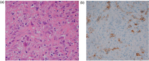
Table 1 Laboratory parameters showing deterioration of coagulation parameters
Fig. 2 Graph showing deterioration of coagulation parameters.
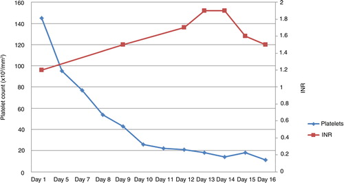
Fig. 3 (a) Liver biopsy showing diffuse infiltration of liver by lymphoma cells with disruption of hepatic architecture and necrosis. (b) Liver biopsy showing hepatocellular necrosis and malignant infiltrate of lymphoma cells.