Figures & data
Figure 1. First scan: axial CT scan with 40 cc intravenous contrast.
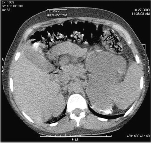
Figure 2. Second scan: 24 hours. After first scan. Arterial phase axial CT scan with 100 cc I.V. contrast.
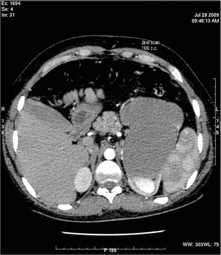
Figure 3. Third scan: additional 100 cc I.V. contrast after second scan.
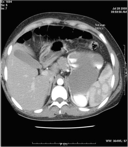
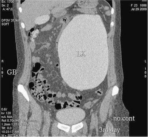
Figure 4. 48 hours delayed scan shows contrast in gall bladder and colon besides hydronephrotic left kidney (marked with oval).
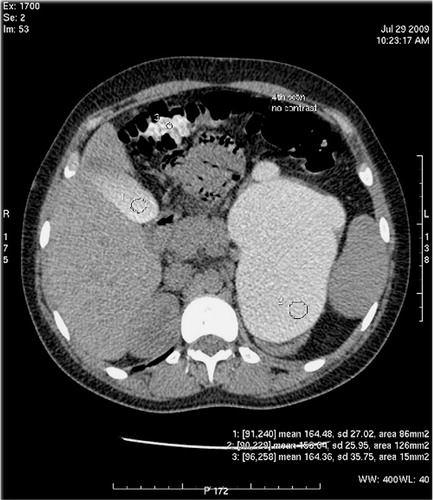
Figure 5. Coronal reformat of delayed scan showing opacified gall bladder and ascending colon besides opacified hydronephrotic left kidney.