Figures & data
Figure 1. Nature of retained foreign bodies.
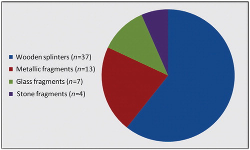
Figure 2. Age distribution (in years).
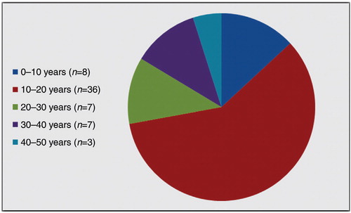
Table 1. Occupational status of the patients
Table 2. Presenting features of missed foreign bodies
Table 3. Time elapsed since possible penetration of foreign body
Figure 3. Management of injuries resulting in missed foreign bodies.
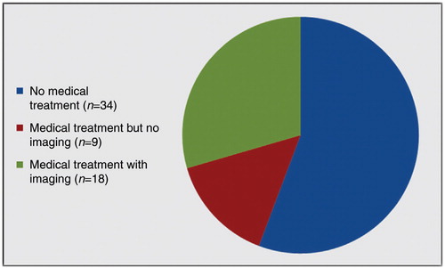
Figure 4. (A) Plain radiograph showing a retained glass piece (arrow). (B) Plain radiograph showing a retained sewing needle. (C) Plain radiograph showing a radio-opaque pellet along proximal phalanx of index finger (arrow). (D) Ultrasonogram revealing echogenic wooden splinters (arrow).
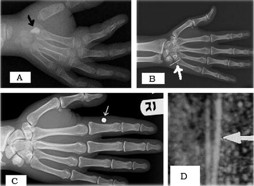
Table 4. Diagnostic modalities used for localizing missed foreign bodies