Figures & data
Fig. 1 Map of the study site. (a) Location of Potter Cove on King George Island (Isla 25 de Mayo), South Shetland Islands. (b) Sampling zone.
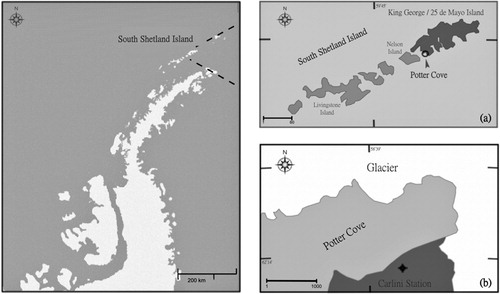
Fig. 2 External morphology of Malacobelemnon daytoni. (a) Tips of colonies showing axial rod. (b) Detail of M. daytoni tips showing the exposed axial rod. (c) Lower rachis and peduncle. (d) Details of the peduncle. (e) Autozooid in the apical part of the colony with mature oocytes.
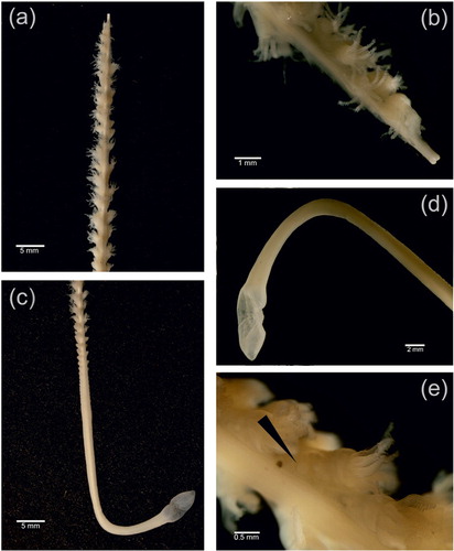
Fig. 3 Relative frequency of oocytes’ diameter in polyps of Malacobelemnon daytoni (100 colonies, n = 5360).
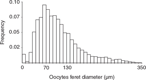
Fig. 4 Relationship between length (mm) and index of fecundity of Malacobelemnon daytoni (number mature oocytes, mm−2).

Fig. 5 Relationship between length of fertile region (rachis) with increasing colony length in Malacobelemnon daytoni.
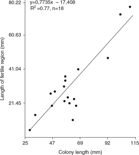
Fig. 6 Malacobelemnon daytoni. (a) Light microscopic section through the longitudinal canal showing early growth oocytes (EO) grouped in cluster (CL). (b) Light microscopic section through the longitudinal canal showing growth oocytes. (c) Mature oocyte in longitudinal canal showing follicle cells (FC).
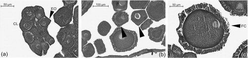
Fig. 7 Mean oocyte stages frequency distribution of Malacobelemnon daytoni in each of the colony sections: basal, middle and apical (n = 671 for basal; n = 3786 for middle and n = 884 for apical) Vertical bars indicate±SD. Letters indicate groups of the a posteriori analysis (p < 0.05).
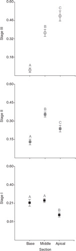
Table 1 Sexual pattern of Pennatulacea, arranged in chronological order of the published source.